Staining
In biochemistry, it involves adding a class-specific (DNA, proteins, lipids, carbohydrates) dye to a substrate to qualify or quantify the presence of a specific compound.
Biological staining is also used to mark cells in flow cytometry, and to flag proteins or nucleic acids in gel electrophoresis.
Light microscopes are used for viewing stained samples at high magnification, typically using bright-field or epi-fluorescence illumination.
Combined with specific protocols for fixation and sample preparation, scientists and physicians can use these standard techniques as consistent, repeatable diagnostic tools.
The liquid is added to the slide before the addition of the organism and a coverslip is placed over the specimen in the water and stain to help contain it within the field of view.
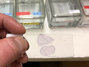
Pieces of tissue may be embedded in paraffin wax to increase their mechanical strength and stability and to make them easier to cut into thin slices.
Becker's method Fontana's mordant(5%Tannic acid) Permeabilization involves treatment of cells with (usually) a mild surfactant.
This means that samples of the manufacturer's batch have been tested by an independent body, the Biological Stain Commission (BSC), and found to meet or exceed certain standards of purity, dye content and performance in staining techniques ensuring more accurately performed experiments and more reliable results.
This can be achieved by smearing the sample onto the slide and then applying nigrosin (a black synthetic dye) or India ink (an aqueous suspension of carbon particles).
After drying, the microorganisms may be viewed in bright field microscopy as lighter inclusions well-contrasted against the dark environment surrounding them.
shapes and arrangements into thin film Gram negative appears pink in color Non acid fast: Blue Vegetative cells: Red A: Hiss method (Positive technique) B: Manevals's technique (Negative) Bacterial suspension smeared along with Congo red and the Maneval's stain is applied Bacteria: Purple capsule, bacterial cell, stands out against dark background Cytoplasm- colorless Cytoplasm: Light pink Cytoplasm: Green Gram staining is used to determine gram status to classifying bacteria broadly based on the composition of their cell wall.
Gram status, helps divide specimens of bacteria into two groups, generally representative of their underlying phylogeny.
This characteristic, in combination with other techniques makes it a useful tool in clinical microbiology laboratories, where it can be important in early selection of appropriate antibiotics.
Due to the presence of higher lipid content, after alcohol-treatment, the porosity of the cell wall increases, hence the CVI complex (crystal violet – iodine) can pass through.
Through the use of malachite green and a diluted ratio of carbol fuchsin, fixing bacteria in osmic acid was a great way to ensure no blending of dyes.
This revision included substitution of carbol fuchsin with aqueous Safranin paired with a newly diluted 5% formula of malachite green.
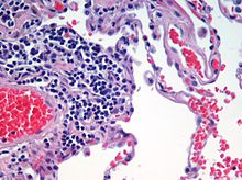
In a skillfully made H&E preparation the red blood cells are almost orange, and collagen and cytoplasm (especially muscle) acquire different shades of pink.
Due to the high volume of carbohydrates within the cell wall of hyphae and yeast forms of fungi, the Periodic acid -Schiff stain can help locate these species inside tissue samples of the human body.
The recipe has evolved from Masson's original technique for different specific applications, but all are well-suited to distinguish cells from surrounding connective tissue.
Acridine orange (AO) is a nucleic acid selective fluorescent cationic dye useful for cell cycle determination.
Some complementing stains used alongside Bismark brown are Hematoxylin and Toluidine blue which provide better contrast within the histology sample.
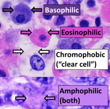
[18] Eosin is most often used as a counterstain to haematoxylin, imparting a pink or red colour to cytoplasmic material, cell membranes, and some extracellular structures.
Consequently, ethidium bromide is often used as a marker for apoptosis in cells populations and to locate bands of DNA in gel electrophoresis.
Hoechst 33258 contains a terminal hydroxyl group and is thus more soluble in aqueous solution, however this characteristics reduces its ability to penetrate the plasma membrane.
When starch is mixed with iodine in solution, an intensely dark blue colour develops, representing a starch/iodine complex.
Used with common vinegar (acetic acid), Lugol's solution is used to identify pre-cancerous and cancerous changes in cervical and vaginal tissues during "Pap smear" follow up examinations in preparation for biopsy.
The acetic acid causes the abnormal cells to blanch white, while the normal tissues stain a mahogany brown from the iodine.
Methyl green is used commonly with bright-field, as well as fluorescence microscopes [20] to dye the chromatin of cells so that they are more easily viewed.
It dissolves in fats, and is reduced by organic materials to elemental osmium, an easily visible black substance.
[26]Phosphotungstic acid is a common negative stain for viruses, nerves, polysaccharides, and other biological tissue materials.