Subepithelial connective tissue graft
[1] Currently, it is generally used to obtain root coverage following gingival recession, which was a later development by Burt Langer in the early 1980s.
Because the connective tissue for the graft is transplanted without the superficial epithelium from the donor site, it is termed subepithelial.
[1] Others, including Broome and Taggert[4] and Donn[5] also described the use of SECT grafts for increasing the zone of keratinized tissue.
Of the various ways of preparing the graft recipient site, Edel described using two vertical incisions, mesial and distal to the teeth at which the zone of keratinized tissue was intended to be widened.
Langer later described the SECT as a method by which to augment concavities and irregularities of the alveolar ridge following traumatic extractions, advanced periodontitis or developmental defects.
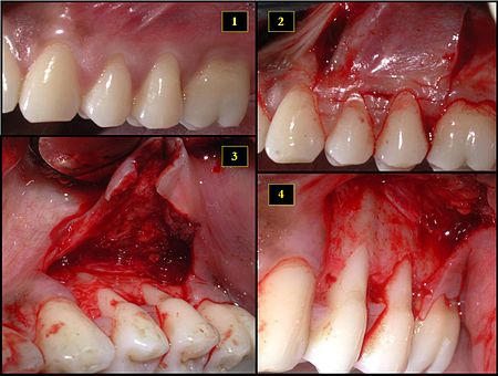
- Recipient site exhibits gingival recession on both premolars and first molar (molar recession is not an esthetic issue and will not be treated)
- Incisions prior to flap reflection
- Full thickness flap elevated
- Another viewpoint of the flapped recipient site

- Ipsilateral palatal mucosa serving as the donor site
- The retrieved connective tissue, approximately 25 × 6 mm in dimension
- Connective tissue placed at recipient site
- Recipient site flap coronally advanced and sutured to entirely cover the graft