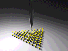
Tip-enhanced Raman spectroscopy
TERS overcomes these limitations as the Raman spectrum obtained originates primarily from the molecules within a few tens of nanometers of the tip.
Although the antennas' electric near-field distributions are commonly understood to determine the spatial resolution, recent experiments showing subnanometer-resolved optical images put this understanding into question.
[2] This is because such images enter a regime in which classical electrodynamical descriptions might no longer be applicable and quantum plasmonic[5] and atomistic[6] effects could become relevant.
In the case of STM-TERS, only side and top illumination configurations can be applied, since the substrate is required to be conductive, therefore typically being non-transparent.
[15][16][17][18] In 2019, the Ara Apkarian group at the Center for Chemistry at the Space-Time Limit, University of California, Irvine imaged vibrational normal modes of single porphyrin molecules using TERS.