Tissue typing
During tissue typing, an individual's human leukocyte antigens (HLA) are identified.
[3] More specifically, HLA mismatches between organ donors and recipients can lead to the development of anti-HLA donor-specific antibodies (DSAs).
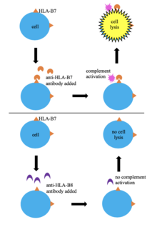
In this technique, a donor's blood cells are HLA typed by mixing them with serum containing anti-HLA antibodies.
This allows for identification of the cells' HLA based indirectly on the specificity of the known antibodies in the serum.
In this method, an unknown HLA sample is mixed with a reference allele and run in a gel by electrophoresis.