Vertebra
The upper and lower surfaces of the vertebra body give attachment to the intervertebral discs.
Vertebrae articulate with each other to give strength and flexibility to the spinal column, and the shape at their back and front aspects determines the range of movement.
Along the length of the spine, the vertebrae change to accommodate different needs related to stress and mobility.
The upper and lower surfaces of the body of the vertebra are flattened and rough in order to give attachment to the intervertebral discs.
These surfaces are the vertebral endplates which are in direct contact with the intervertebral discs and form the joint.
[11][12] There are superior and inferior articular facet joints on each side of the vertebra, which serve to restrict the range of movement possible.
These facets are joined by a thin portion of the vertebral arch called the pars interarticularis.
Cervical vertebrae possess transverse foramina to allow for the vertebral arteries to pass through on their way to the foramen magnum to end in the circle of Willis.
This is an opening on each of the transverse processes which gives passage to the vertebral artery and vein and a sympathetic nerve plexus.
Some rotation can occur between the thoracic vertebrae, but their connection with the rib cage prevents much flexion or other movement.
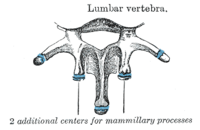
As the vertebrae progress down the spine they increase in size to match up with the adjoining lumbar section.
The discs between these vertebrae create a natural lumbar lordosis (a spinal curvature that is concave posteriorly).
[citation needed] This is due to the difference in thickness between the front and back parts of the intervertebral discs.
The term lumbosacral is often used to refer to the lumbar and sacral vertebrae together, and sometimes includes their surrounding areas.
There are five sacral vertebrae (S1–S5) which are fused in maturity, into one large bone, the sacrum, with no intervertebral discs.
[18] The sacrum with the ilium forms a sacroiliac joint on each side of the pelvis, which articulates with the hips.
Other cells move distally to the costal processes of thoracic vertebrae to form the ribs.
Wedge-shaped vertebrae, called hemivertebrae can cause an angle to form in the spine which can result in the spinal curvature diseases of kyphosis, scoliosis and lordosis.
A laminectomy is a surgical operation to remove the laminae in order to access the spinal canal.
The vertebral pedicle is often used as a radiographic marker and entry point in vertebroplasty, kyphoplasty, and spinal fusion procedures.
It is a bony bridge found on the first cervical vertebra, the atlas where it covers the groove for the vertebral artery.
[19] Because of the different types of locomotion and support needed between the aquatic and other vertebrates, the vertebrae between them show the most variation, though basic features are shared.
The spinous processes which are backward extending are directed upward in animals without an erect stance.
Spinous processes are exaggerated in some animals, such as the extinct Dimetrodon and Spinosaurus, where they form a sailback or finback.
[25] This type of connection permits a wide range of motion in most directions, while still protecting the underlying nerve cord.
The central point of rotation is located at the midline of each centrum, and therefore flexion of the muscle surrounding the vertebral column does not lead to an opening between vertebrae.
In many groups, such as lizards and saurischian dinosaurs, the cervical ribs are large; in birds, they are small and completely fused to the vertebrae.
In the whale, the cervical vertebrae are typically fused, an adaptation trading flexibility for stability during swimming.
In humans and other tailless primates, they are called the coccygeal vertebrae, number from three to five and are fused into the coccyx.
[36] This article incorporates text in the public domain from page 96 of the 20th edition of Gray's Anatomy (1918)