Visual field
[6] The corresponding concept for optical instruments and image sensors is the field of view (FOV).
In humans and animals, the FOV refers to the area visible when eye movements – if possible for the species – are allowed.
[13] The nose is situated in the field of view of both eyes, but due to later processing carried out in the brain, it is not noticed during normal visual tasks.
This may be kinetic, where spots of light are shown on the white interior of a half sphere and slowly moved inwards until the observer sees them, or static, where the light spots are flashed at varying intensities at fixed locations in the sphere until detected by the subject.
Another method is to use a campimeter, a small device with a flat screen designed to measure the central visual field.
Another method is for the practitioner to hold up one, two, or five fingers in the four quadrants and center of a patient's visual field (with the other eye covered).
Visual field loss may occur due to many disease or disorders of the eye, optic nerve, or brain.
Most of the early glaucomatous changes are seen within the central visual field, mainly in Bjerrum's area, 10°-20° from fixation.

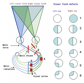
From top to bottom:
1. Complete loss of vision in the right eye
2. Bitemporal hemianopia
3. Homonymous hemianopia
4. Quadrantanopia
5. & 6. Quadrantanopia with macular sparing