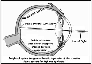
Peripheral vision
[1] The fovea is a cone-shaped depression in the central retina measuring 1.5 mm in diameter, corresponding[5] to 5° of the visual field.
When viewed through the pupil, as in an eye exam (using an ophthalmoscope or retinal photography), only the central portion of the fovea may be visible.
This corresponds to using the foveal avascular zone (FAZ) with a diameter of 0.5 mm representing 1.5° of the visual field (although often idealized as perfect circles, the central structures of the retina tend to be irregular ovals).
[19][12] The term is familiar in the general public through the widespread macular degeneration (AMD) at older age, where central vision is lost.
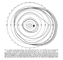
[20] A dividing line between near and mid peripheral vision at 30° radius can be based on several features of visual performance.
[24] In dark-adapted vision, light sensitivity corresponds to rod density,[citation needed] which peaks just at 18°.
From 18° away from the center, rod density declines more gradually, in a curve with distinct inflection points resulting in two humps.
[citation needed] The distinctions between foveal (sometimes also called central) and peripheral vision are reflected in subtle physiological and anatomical differences in the visual cortex.
It has been suggested that these areas are important for fast reactions to visual stimuli in the periphery, and monitoring body position relative to gravity.
[40] The main functions of peripheral vision are:[38] When viewed at large angles, the iris and pupil appear to be rotated toward the viewer due to the optical refraction in the cornea.
The retina extends farthest in the superior-nasal 45° quadrant (in the direction from the pupil to the bridge of the nose) with the greatest extent of the visual field in the opposite direction, the inferior temporal 45° quadrant (from the pupil of either eye towards the bottom of the nearest ear).
Vision at this extreme part of the visual field is thought to be possibly concerned with threat detection, measuring optical flow, color constancy, or circadian rhythm.