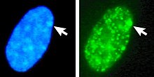
Barr body
In species with XY sex-determination (including humans), females typically have two X chromosomes,[2] and one is rendered inactive in a process called lyonization.
[6] The center contains twelve genes, seven of which code for proteins, five for untranslated RNAs, of which only two are known to play an active role in the X inactivation process, Xist and Tsix.
[3] Variations in Xi frequency have been reported with age, pregnancy, the use of oral contraceptives, fluctuations in menstrual cycle and neoplasia.
[12] One study showed that the frequency of Barr bodies in breast carcinoma were significantly lower than in healthy controls, indicating reactivation of these once inactivated X chromosomes.
Their detection in ancient samples provides a powerful tool for gender identification in extinct species, offering insights into population dynamics, biology, and evolution.
Recent advancements in histological and genomic techniques have made it possible to observe Barr bodies in ancient remains, including fossilized bones and tissues: In a notable example, Barr bodies were detected in osteocytes from ancient mammalian remains, demonstrating the potential of this approach for studying gender in extinct populations.