Bicornuate uterus
[2] Bicornuate uterus is typically classified based on whether or not the division extends to the external cervical os.
[2] There is a continuous range of the degree and location of the fusion of the paramesonephric ducts, and existence of a spectrum, rather than a fixed number of types corresponding to strict medical definitions.
Two processes that occur during the embryonic development of the paramesonephric ducts — fusion and reabsorption — can be affected to different degrees.
[1] Women with a bicornuate uterus are at an increased risk of recurrent miscarriage,[2][10] preterm birth,[2][11] malpresentation,[2][12] disruptions to fetal growth,[13] premature rupture of membranes, placenta previa and retained placenta (which can lead to postpartum hemorrhage).
[14] Fetuses developing in bicornuate uteri are more likely to present breech or transverse, with the fetal head in one horn and the feet in the other.
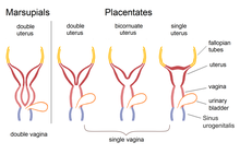
In insectivores, certain species of primates i.e. lemurs, furthermore in carnivores, and in whales the uterus bicornis a part of their healthy morphological structures.
If the presence of a uterus bicornis is not yet known and there is clinical suspicion of a carcinoma, which cannot be confirmed by the histological findings, sonography and magnetic resonance imaging provide diagnostic possibilities, as the cause may lie in a second uterine horn.