Obstetric ultrasonography
[2] Research shows that routine obstetric ultrasound before 24 weeks' gestational age can significantly reduce the risk of failing to recognize multiple gestations and can improve pregnancy dating to reduce the risk of labor induction for post-dates pregnancy.
[3] Below are useful terms on ultrasound:[4] In normal state, each body tissue type, such as liver, spleen or kidney, has a unique echogenicity.
[6] While 3D is popular with parents desiring a prenatal photograph as a keepsake,[7] both 2D and 3D are discouraged by the FDA for non-medical use,[8] but there are no definitive studies linking ultrasound to any adverse medical effects.
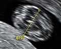
[13] In the first trimester, a standard ultrasound examination typically includes:[12] In the second trimester, a standard ultrasound exam typically includes:[12] Gestational age is usually determined by the date of the woman's last menstrual period, and assuming ovulation occurred on day fourteen of the menstrual cycle.
Sometimes a woman may be uncertain of the date of her last menstrual period, or there may be reason to suspect ovulation occurred significantly earlier or later than the fourteenth day of her cycle.
[23] Current evidence indicates that diagnostic ultrasound is safe for the unborn child, unlike radiographs, which employ ionizing radiation.
Randomized controlled trials have followed children up to ages 8–9, with no significant differences in vision, hearing, school performance, dyslexia, or speech and neurologic development by exposure to ultrasound.
[24] Several randomized controlled trials have reported no association between Doppler exposure and birth weight, Apgar scores, and perinatal mortality.
One randomized controlled trial, however, came to the result of a higher perinatal death rate of normally formed infants born after 24 weeks exposed to Doppler ultrasonography (RR 3.95, 95% CI 1.32–11.77), but this was not a primary outcome of the study, and has been speculated to be due to chance rather than a harmful effect of Doppler itself.
[27] The American Institute of Ultrasound in Medicine recommends spectral Doppler only if M-mode sonography is unsuccessful, and even then only briefly, due to the acoustic intensity delivered to the fetus.
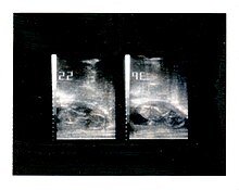
[31][32] In 1962, after about two years of work, Joseph Holmes, William Wright, and Ralph Meyerdirk developed the first compound contact B-mode scanner.
Wright and Meyerdirk left the university to form Physionic Engineering Inc., which launched the first commercial hand-held articulated arm compound contact B-mode scanner in 1963.
[citation needed] In March and April 2015, a post by a pregnant woman named Jen Martin (née Cardinal) and her husband to YouTube, which had been viewed at least 2 million times and had many likes, showed the 14-week-old fetus clapping repeatedly to the song, sung by the parents, "If You're Happy And You Know It".
[37] Ethnographic research concerned with the use of ultrasound technology in monitoring pregnancy can show us how it has changed the embodied experience of expecting mothers around the globe.
[37] Recent studies have stressed the importance of framing "reproductive health matters cross-culturally", particularly when understanding the "new phenomenon" of "the proliferation of ultrasound imaging" in developing countries.
[38] Therefore, although women, particularly in Asian countries, "express intense uncertainties regarding the safety and credibility of this technology", it is overused for its "immediate reassurance".