Cathodoluminescence
Cathodoluminescence is an optical and electromagnetic phenomenon in which electrons impacting on a luminescent material such as a phosphor, cause the emission of photons which may have wavelengths in the visible spectrum.
A familiar example is the generation of light by an electron beam scanning the phosphor-coated inner surface of the screen of a television that uses a cathode-ray tube.
In terms of band structure, classical semiconductors, insulators, ceramics, gemstones, minerals, and glasses can be treated the same way.
It is designed to image the luminescence characteristics of polished thin sections of solids irradiated by an electron beam.
Using a cathodoluminescence microscope, structures within crystals or fabrics can be made visible which cannot be seen in normal light conditions.
CL color and intensity are dependent on the characteristics of the sample and on the working conditions of the electron gun.
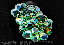
The advantage of a hot cathode is the precisely controllable high beam intensity allowing to stimulate the emission of light even on weakly luminescing materials (e.g. quartz – see picture).
From there, a fiber optic will transfer the light out of the microscope where it is separated into its component wavelengths by a monochromator and is then detected with a photomultiplier tube.
Instead, by measuring the wavelength dependence for a fixed point or a certain area, the spectral characteristics can be recorded (cathodoluminescence spectroscopy).
Furthermore, if the photomultiplier tube is replaced with a CCD camera, an entire spectrum can be measured at each point of a map (hyperspectral imaging).
Recently, cathodoluminescence performed in electron microscopes is also being used to study surface plasmon resonances in metallic nanoparticles.