Chromoendoscopy
Chromoendoscopy is a medical procedure wherein dyes (often the same stains used in histology) are instilled into the gastrointestinal tract at the time of visualization with fibre-optic endoscopy.
[citation needed] Reactive stains undergo an observable change due to a chemical process related to the function of the gastrointestinal tract.
[1] A Cochrane review updated in 2016 found strong evidence that chromoscopy enhances the detection of cancerous tumours in the colon and rectum when compared to plain colonoscopy.
[1] The stains are typically applied to the mucosal lining of the gastrointestinal tract using a device known as a spray catheter that is inserted into the endoscope.
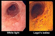
Depending on the reason for which chromoendoscopy is being performed, the dye can be sprayed directly on a suspected abnormality (such as for identification of squamous cell carcinoma) or diffusely to the mucosa (such as for screening for dysplasia in ulcerative colitis[1] When the stain is applied diffusely in the colon, a complete colonoscopy is performed with the instrument lying in the cecum.
Narrow-band imaging is a technique where ambient light of a blue and green wavelengths are used to highlight detail, particularly of vascular structures.
[6] Other techniques that can enhance detail of mucosa include confocal microscopy, magnification endoscopy and optical coherence tomography.