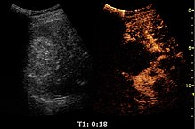
Contrast-enhanced ultrasound
Ultrasound contrast agents rely on the different ways in which sound waves are reflected from interfaces between substances.
Although colour Doppler can be used to detect abnormal flows between the chambers of the heart (e.g., persistent (patent) foramen ovale), it has a limited sensitivity.
When specifically looking for a defect such as this, small air bubbles can be used as a contrast medium and injected intravenously, where they travel to the right side of the heart.
[citation needed] Regardless of the shell or gas core composition, microbubble size is fairly uniform.
[11] In addition to the PEG layer, the shell is modified with molecules that allow for the attachment of ligands that bind certain receptors.
Humanizing antibodies is an expensive and time-intensive process, so it would be ideal to find an alternative source of ligands, such as synthetically manufactured targeting peptides that perform the same function, but without the immune issues.
[citation needed] There are two forms of contrast-enhanced ultrasound, untargeted (used in the clinic today) and targeted (under preclinical development).
Untargeted microbubbles, such as the aforementioned SonoVue, Optison, or Levovist, are injected intravenously into the systemic circulation in a small bolus.
Microbubbles targeted with ligands that bind certain molecular markers that are expressed by the area of imaging interest are still injected systemically in a small bolus.
Microbubbles theoretically travel through the circulatory system, eventually finding their respective targets and binding specifically.
If a sufficient number of microbubbles have bound in the area, their compressible gas cores oscillate in response to the high frequency sonic energy field, as described in the ultrasound article.
The ultrasound system converts the strong echogenicity into a contrast-enhanced image of the area of interest, revealing the location of the bound microbubbles.