Cryptomonad
The two groups are united under three shared morphological characteristics: presence of a periplast, ejectisomes with secondary scroll, and mitochondrial cristae with flat tubules.
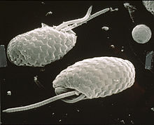
[8] Cryptomonads are distinguished by the presence of characteristic extrusomes called ejectosomes, which consist of two connected spiral ribbons held under tension.
Large ejectosomes, visible under the light microscope, are associated with the pocket; smaller ones occur underneath the periplast, the cryptophyte-specific cell surrounding.
In the case of Rhodomonas, the crystal structure has been determined to 1.63 Å;[14] and it has been shown that the alpha subunit bears no relation to any other known phycobiliprotein.
Some Cryptomonas species may also form immotile microbial cysts—resting stages with rigid cell walls to survive unfavorable conditions.
Cryptomonad flagella are inserted parallel to one another, and are covered by bipartite hairs called mastigonemes, formed within the endoplasmic reticulum and transported to the cell surface.

- Anterior flagellum ( mastigonemes on both faces)
- Posterior flagellum (mastigonemes on one face)
- Contractile vacuole , regulates the quantity of water inside a cell
- Vestibulum
- Basal bodies
- Gullet (furrow or crypt)
- Mitochondrion , creates ATP (energy) for the cell
- Maupa's bodies
- Ejectisomes
- Starch granule
- Golgi apparatus , packages proteins
- Nucleomorph , a small, vestigial eukaryotic nucleus
- Pyrenoid , center of carbon fixation
- Periplastidial compartment
- Thylakoid , site of the light-dependent reactions of photosynthesis
- Plastid membranes (4, secondary)
- Nucleus
- Nucleolus
- Lipid globules