Dermatophyte
[2] Traditionally, these anamorphic (asexual or imperfect fungi) mold genera are: Microsporum, Epidermophyton and Trichophyton.
[4] Dermatophytes cause infections of the skin, hair, and nails, obtaining nutrients from keratinized material.
Colonies of dermatophytes are usually restricted to the nonliving cornified layer of the epidermis because of their inability to penetrate the viable tissue of an immunocompetent host.
[8] Although symptoms can be barely noticeable in some cases, dermatophytoses can produce "chronic progressive eruptions that last months or years, causing considerable discomfort and disfiguration.
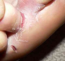
[3] Later stages of tinea pedis might include hyperkeratosis (thickened skin) of the soles, as well as bacterial infection (by streptococcus and staphylococcus) or cellulitis due to fissures developing between the toes.
[3][11] Another implication of tinea pedis, especially for older adults or those with vascular disease, diabetes mellitus, or nail trauma, is onychomycosis of the toenails.
[3] Lesions appear as round, red, scaly, patches with well-defined, raised edges, often with a central clearing and very itchy (usually on trunk, limbs, and also in other body parts).
Trichophyton rubrum is also a very common cause of favus, a form of tinea capitis in which crusts are seen on the scalp.
Infected hair shafts are broken off just at the base, leaving a black dot just under the surface of the skin, and alopecia can result.
[14] See Onychomycosis Ringworm infections modified by corticosteroids, systemic or topical, prescribed for some pre-existing pathology or given mistakenly for the treatment of misdiagnosed tinea.
[3] The fungi spread in a centrifugal pattern in the stratum corneum, which is the outermost keratinized layer of the skin.
[8] Dermatophytoses rarely cause serious illness, as the fungi infection tends to be limited to the superficial skin.
[9] The infection tends to self-resolve so long as the fungal growth does not exceed inflammatory response and desquamation rate is sufficient.
[8] Infection may become chronic and widespread if the host has a compromised immune system and is receiving treatment that reduces T-lymphocyte function.
[8] However, "the clinical manifestations of these infections are largely due to delayed-type hypersensitivity responses to these agents rather than from direct effects of the fungus on the host.
[3] However, a confirmatory rapid in-office test can also be conducted, which entails using a scalpel to scrape off a lesion sample from the nail, skin, or scalp and transferring it to a slide.
Potassium hydroxide (KOH) is added to the slide and the sample is examined with a microscope to determine presence of hyphae.
[9] Additionally, a Wood's lamp examination (ultraviolet light) may be used to diagnose specific dermatophytes that fluoresce.
Microscopic morphology of the micro- and macroconidia is the most reliable identification character, but both good slide preparation and stimulation of sporulation in some strains are needed.
[9] Dermatophytes are transmitted by direct contact with an infected host (human or animal)[3] or by direct or indirect contact with infected shed skin or hair in fomites such as clothing, combs, hair brushes, theatre seats, caps, furniture, bed linens, shoes,[16] socks,[16] towels, hotel rugs, sauna, bathhouse, and locker room floors.
Adaptation to growth on humans by most geophilic species resulted in diminished loss of sporulation, sexuality, and other soil-associated characteristics.
Dermatophytes are classified as anthropophilic (humans), zoophilic (animals) or geophilic (soil) according to their normal habitat.
[17] In heterothallic species, interaction of two individuals with compatible mating types are required in order for sexual reproduction to occur.
In contrast, homothallic fungi are self-fertile and can complete a sexual cycle without a partner of opposite mating type.
Tinea capitis (scalp) must be treated orally, as the medication must be present deep in the hair follicles to eradicate the fungus.