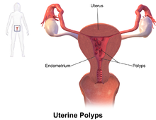
Endometrial polyp
[5] No definitive cause of endometrial polyps is known, but they appear to be affected by hormone levels and grow in response to circulating estrogen.
[3] Risk factors include obesity, high blood pressure and a history of cervical polyps.
[3] Taking tamoxifen or hormone replacement therapy can also increase the risk of uterine polyps.
[3][8] The use of an intrauterine system containing levonorgestrel in women taking tamoxifen may reduce the incidence of polyps.
[9] Endometrial polyps can be detected by vaginal ultrasound (sonohysterography), hysteroscopy and dilation and curettage.
[7] The polyps consist of dense, fibrous tissue (stroma), blood vessels and glandlike spaces lined with endometrial epithelium.
To reduce this risk, the uterus can be first explored using grasping forceps at the beginning of the curettage procedure.
[7] Hysteroscopy involves visualising the endometrium (inner lining of the uterus) and polyp with a camera inserted through the cervix.