Eukaryotic DNA replication
In eukaryotes, the vast majority of DNA synthesis occurs during S phase of the cell cycle, and the entire genome must be unwound and duplicated to form two daughter copies.
[8][14] Generally, the function and size of the ORC subunits are conserved throughout many eukaryotic genomes with the difference being their diverged DNA binding sites.
The most widely studied origin recognition complex is that of Saccharomyces cerevisiae or yeast which is known to bind to the autonomously replicating sequence (ARS).
[15] The S. cerevisiae ORC interacts specifically with both the A and B1 elements of yeast origins of replication, spanning a region of 30 base pairs.
All five subunits contact the sugar phosphate backbone at multiple points of the A element to form a tight grip without base specificity.
[16] The bending of origin DNA by ORC appears to be evolutionarily conserved suggesting that it may be required for the Mcm2-7 complex loading mechanism.
[16][19] When the ORC binds to DNA at replication origins, it serves as a scaffold for the assembly of other key initiation factors of the pre-replicative complex.
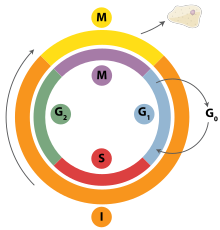
[20] This pre-replicative complex assembly during the G1 stage of the cell cycle is required prior to the activation of DNA replication during the S phase.
[31] The minichromosome maintenance (Mcm) proteins were named after a genetic screen for DNA replication initiation mutants in S. cerevisiae that affect plasmid stability in an ARS-specific manner.
[58] The Xenopus nucleus-free system also demonstrates that DNA unwinding and tight RPA binding to chromatin occurs only in the presence of Cdc45.
In eukaryotic cells, a small amount of the DNA segment immediately upstream of the RNA primer is also displaced, creating a flap structure.
In lagging strand synthesis, the movement of DNA polymerase in the opposite direction of the replication fork requires the use of multiple RNA primers.
This generates an RNA-DNA single strand flap, which must be cleaved, and the nick between the two Okazaki fragments must be sealed by DNA ligase I.
[95] It has been proposed that this iterative process is preferable to the cell because it is tightly regulated and does not generate large flaps that need to be excised.
Electron microscopy studies indicate that nucleosome loading on the lagging strand occurs very close to the site of synthesis.
[3] Importantly, this priming action occurs at replication initiation at origins to begin leading-strand synthesis and also at the 5' end of each Okazaki fragment on the lagging strand.
To thwart these problems, the eukaryotic replisome contains specialized proteins that are designed to regulate the helicase activity ahead of the replication fork.
These proteins also provide docking sites for physical interaction between helicases and polymerases, thereby ensuring that duplex unwinding is coupled with DNA synthesis.
[113] The importance of this direct link between the helicase and the leading-strand polymerase is underscored by results in cultured human cells, where Mrc1/Claspin is required for efficient replication fork progression.
This constant initiation of Okazaki fragment synthesis requires repeated PCNA loading for efficient DNA replication.
[119][120] The PCNA homotrimer is opened by RFC by ATP hydrolysis and is then loaded onto DNA in the proper orientation to facilitate its association with the polymerase.
Due to the fact that an RNA primer is required for initiation of DNA synthesis, the lagging strand is at a disadvantage in replicating the entire chromosome.
The RNA component of telomerase anneals to the single stranded 3' end of the template DNA and contains 1.5 copies of the telomeric sequence.
This high level of CDK activity is responsible for initiating DNA replication as well as inhibiting new pre-RC complex formation.
[2] Geminin first appears in S-phase and is degraded at the metaphase-anaphase transition, possibly through ubiquination by anaphase promoting complex (APC).
Checkpoint proteins are also involved in some DNA repair pathways, while they stabilize the structure of the replication fork to prevent further damage.
This mechanism prevents continued DNA synthesis and is required for the protection of the genome in the presence of replication stress and potential genotoxic conditions.
[145] CAF-1 contains a PCNA-binding motif, called a PIP-box, that allows CAF-1 to associate with the replisome through PCNA and is able to deposit histone H3-H4 dimers onto newly synthesized DNA.
[149] Electron microscopy studies show that this occurs very quickly, as nucleosomes can be observed forming just a few hundred base pairs after the replication fork.
[152] These enzymes work to promote MiDAS, operating outside of ATR, BRCA2, and RAD51 which are necessary to prevent replication stress at CFS loci throughout S phase.