Ff phages
[1][2][3][4][5][6][7] The virion (virus particle) is a flexible filament measuring about 6 by 900 nm, comprising a cylindrical protein tube protecting a single-stranded circular DNA molecule at its core.
George Smith and Greg Winter used f1 and fd for their work on phage display for which they were awarded a share of the 2018 Nobel Prize in Chemistry.
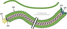
Several thousand copies of a small (50 amino-acid residues) elongated alpha-helical major coat protein subunit (the product of gene 8, or p8) in an overlapping shingle-like array form a hollow cylinder enclosing the circular single-stranded DNA genome.
The shingle-like arrangement places the acidic residues of p8 near the outside surface of the cylinder, where they cause the virus particle to be negatively-charged; non-polar regions near non-polar regions of neighbouring p8 subunits, where non-polar interactions contribute to a notable physical stability of the virus particle; and basic residues near the centre of the cylinder, where they interact with the negatively-charged DNA phosphates at the core of the virion.
In particular, the series of fd and Pf1 virion structures deposited in the PDB over decades illustrate the improvements in methods for fiber diffraction data collection and computational analysis.
[1][19][20] As the tip of the pilus bearing p3 approaches the cell wall, the N1 domain of p3 interacts with the bacterial TolQRA protein to complete infection and release the genome into the cytoplasm of the host.
The complementary strand of the RF is the transcription template for phage coded proteins, especially p2 and p10, which are necessary for further DNA replication.
When a circle is complete, the covalently linked p2 cuts the displaced viral strand at the junction between the old and newly synthesized DNA and re-ligates the two ends and liberates p2.
The extrusion process picks up the p7 and p9 proteins which form the outer tip of the progeny phage.
[14] Interaction of the double-stranded packaging DNA signal with the p1-thioredoxin complex at the host inner membrane triggers the formation of a pore.
The traditional “tadpole” or isometric shaped-phage, on the other hand, which have a limited-sized capsid, cannot be so easily used to encapsidate a larger DNA molecule.
The reverse of this approach is to insert DNA coding for antibodies into gene 3 and detect their presence by appropriate antigens.
[35][36][37] Ff phages have been engineered for applications such as remediation, electrochemical, photovoltaic, catalytic, sensing and digital memory devices, especially by Angela Belcher and colleagues.