Fluorescent tag
The fluorophore selectively binds to a specific region or functional group on the target molecule and can be attached chemically or biologically.
The most commonly labelled molecules are antibodies, proteins, amino acids and peptides which are then used as specific probes for detection of a particular target.
[2] The development of methods to detect and identify biomolecules has been motivated by the ability to improve the study of molecular structure and interactions.
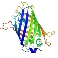
[5] Green fluorescent protein or GFP was discovered by Osamu Shimomura in the 1960s and was developed as a tracer molecule by Douglas Prasher in 1987.
[7] New methods for tracking biomolecules have been developed including the use of colorimetric biosensors, photochromic compounds, biomaterials, and electrochemical sensors.
In this case, amino acids with stable isotopes of either carbon, nitrogen, or hydrogen are incorporated into polypeptide sequences.
A researcher would be able to inspect and get data about the surrounding environment based on what color he or she could see visibly from the biosensor-molecule hybrid species.
They detect changes and measure current between a probed metal electrode and an electrolyte containing the target analyte.
For example, one technique using electrochemical sensing includes slowly raising the voltage causing chemical species at the electrode to be oxidized or reduced.
Cell current vs voltage is plotted which can ultimately identify the quantity of chemical species consumed or produced at the electrode.
[18] For instance, FAST is a variant of photoactive yellow protein which was engineered to bind chemical mimics of the GFP tripeptide chromophore.
Transition metals are used to link specific residues in the tags to site-specific targets such as the N-termini, C-termini, or internal sites within the protein.
Multiple fluorescent dyes that each have a distinct excitation and emission wavelength are bound to a probe which is then hybridized to chromosomes.
A fluorescence microscope can detect the dyes present and send it to a computer that can reveal the karyotype of a cell.
In vivo imaging studies in live animals have been performed for the first time with the use of a monomeric protein derived from the bacterial haloalkane dehalogenase known as the Halo-tag.
[26] Although fluorescent dyes may not have the same sensitivity as radioactive probes, they are able to show real-time activity of molecules in action.
In live cell imaging, fluorescent tags enable movements of proteins and their interactions to be monitored.
[24] Latest advances in methods involving fluorescent tags have led to the visualization of mRNA and its localization within various organisms.
This technique was used to show how the oskar mRNA in the Drosophila embryo localizes to the posterior region of the oocyte.