Forensic serology
Forensic serology is the detection, identification, classification, and study of various bodily fluids such as blood, semen, saliva, and urine, and their relationship to a crime scene.
The most publicized test by crime shows is the Luminol process in which a chemical is sprayed onto a surface where blood is suspected to be.
As with all presumptive tests, this technique can produce false positive results due to metals and strong chemicals, such as bleach, that will also react.
This is a catalytic test that detects the heme group in blood that transports oxygen and carbon dioxide.
[5] A sterile cotton swab is soaked in distilled water and applied to the area of suspected blood to pick up some of the sample.
The Takayama Crystal Assay, which forms a ferro protoporphyrin ring by a reaction between pyridine and the iron atom of the heme group.
[3] Under UV light, semen fluoresces making it visible to investigators to collect samples from a crime scene.
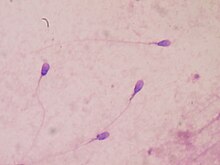
For the christmas tree stain, the sample is extracted with sterile water in order to make a wet mount on a microscope slide.
The sample is then heat-fixed to the slide and stained with nuclear fast red for 15 minutes, then rinsed with deionized water.
[4] This detection technique is based on the activity of the enzyme alpha-amylase which breaks down starches from food into smaller oligosaccharide molecules, starting digestion in the mouth.
If saliva is present, then the alpha-amylase breaks down the starch, creating a clear coloured circle around where the sample was placed.
RSID tests have also been made in order to detect alpha-amylase, but they are not always reliable because there can be a lot of false positives.
MiRNAs are also an ideal target for forensic analysis because they are small compared to other cellular components, so they tend to resist degradation better than other tissue markers, which is important considering that case work samples are not always going to be in pristine condition.
Unlike routine DNA profiling, miRNA amplification requires an extra step before the PCR process.
[15] Once this conversion has happened, the cDNA and the other DNA in the sample can be amplified using PCR and then separated/visualized using a capillary electrophoresis protocol.
Researchers have also added other factors to the loop-mediated isothermal amplification make identification of different body fluids.
Although it has proven to decrease total and hands-on time needed to get a result, there are still kinks to work out before this method is used in many or all forensic labs.