Gamma spectroscopy
When these emissions are detected and analyzed with a spectroscopy system, a gamma-ray energy spectrum can be produced.
Such sources typically produce gamma-ray "line spectra" (i.e., many photons emitted at discrete energies), whereas much higher energies (upwards of 1 TeV) may occur in the continuum spectra observed in astrophysics and elementary particle physics.
Additional components may include signal amplifiers, rate meters, peak position stabilizers, and data handling devices.
With Compton interaction or pair production, a portion of the energy may escape from the detector volume, without being absorbed.
In the MCA, a pulse-shaping amplifier takes the transient voltage signal and reshapes it into a Gaussian or trapezoidal shape.
Additional logic in the MCA then performs pulse-height analysis, sorting the pulses by their height into specific bins, or channels.
[5] A USB sound card can serve as a cheap, consumer off-the-shelf ADC, a technique pioneered by Marek Dolleiser.
Specialized computer software performs pulse-height analysis on the digitized waveform, forming a complete MCA.
[6] Sound cards have high-speed but low-resolution (up to 192 kHz) ADC chips, allowing for reasonable quality for a low-to-medium count rate.
Gamma spectroscopy systems are designed and adjusted to produce symmetrical peaks of the best possible resolution.
The most common figure used to express detector resolution is full width at half maximum (FWHM).
Absolute efficiency values represent the probability that a gamma ray of a specified energy passing through the detector will interact and be detected.
Relative efficiency values greater than one hundred percent can therefore be encountered when working with very large germanium detectors.
The intensity of the light produced is usually proportional to the energy deposited in the crystal by the gamma ray; a well known situation where this relationship fails is the absorption of < 200 keV radiation by intrinsic and doped sodium iodide detectors.
The 662 keV line shown is actually produced by 137mBa, the decay product of 137Cs, which is in secular equilibrium with 137Cs.
The distribution arises because of primary gamma rays undergoing Compton scattering within the crystal: Depending on the scattering angle, the Compton electrons have different energies and hence produce pulses in different energy channels.
Gamma ray reduction techniques are especially useful for small lithium-doped germanium (Ge(Li)) detectors.
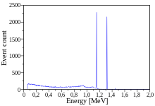
The two gamma lines can be seen well-separated; the peak to the left of channel 200 most likely indicates a strong background radiation source that has not been subtracted.
Because of the poor resolution of NaI-based detectors, they are not suitable for the identification of complicated mixtures of gamma ray-producing materials.
However, a disadvantage is the requirement of cryogenic temperatures for the operation of germanium detectors, typically by cooling with liquid nitrogen.
The detailed shape of backscatter peak structure is influenced by many factors, such as the geometry of the experiment (source geometry, relative position of source, shielding and detector) or the type of the surrounding material (giving rise to different ratios of the cross sections of Photo- and Compton-effect).
The basic principle, however, is as follows: The backscatter peak usually appears wide and occurs at lower than 250 keV.
[11][12] For incident photon energies E larger than two times the rest mass of the electron (1.022 MeV), pair production can occur.
The resulting positron annihilates with one of the surrounding electrons, typically producing two photons with 511 keV.
If a gamma spectrometer is used for identifying samples of unknown composition, its energy scale must be calibrated first.

Ra
, 214
Pb
, and 214
Bi
of the uranium decay chain . This spectrum was taken from a Uranium ore sample from Moab, Utah


Cs
)

Co
); see also a different measurement