Giardia duodenalis
[4][5] G. duodenalis is a non-invasive parasite, that does not spread to other parts of the gastrointestinal tract, but remains confined to the lumen of the small intestine.
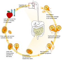
Common symptoms include abdominal pain, nausea, and bloating, along with large, watery, foul-smelling, and greasy stools.
Trophozoites are the replicative stage of the parasite, characterized by a pear-shaped, motile, flagellated cell that survives only in the small intestine of the host.
[8] The infection process in the host begins with the ingestion of cysts, which pass through the stomach into the first part of small intestine, or the duodenum.
Trophozoites cause structural and functional damage to the host epithelial cells, impairing the intestine's ability to absorb nutrients effectively.
[19] The mitosome is a double-membraned organelle, that lacks the enzymatic components required for classic mitochondrial functions, such as ATP synthesis and lipid metabolism.
The metabolic activity of the cyst is significantly lower compared to that of the trophozoites, which allows the parasite to survive for longer periods in harsh conditions, such as in cold environments.
[21] Giardia has common seasonal patterns in the distribution of infection rates with highest peaks in the late summer to early fall.
[23] They may also occur in city reservoirs and persist after water treatment, as the cysts are resistant to conventional water-treatment methods, such as chlorination and ozonolysis.
[citation needed] In addition to waterborne sources, Giardia infections are more commonly found in children compared to adults, this is believed to be due to fecal-oral transmission of the cysts.
7% of children aged 1 to 3 years and 11% of infants and toddlers tested for admission to day-care centers were found to be infected.
[20][24] Giardia duodenalis is common around the world because the parasite resides in bodies of water; typically rivers, lakes, and recreational swimming pools.
[28] Cats can be cured easily, and lambs usually simply lose weight, but in calves, the parasites can be fatal and often are not responsive to antibiotics or electrolytes.
Kennels and areas used for exercise should be considered contaminated for at least one month after dogs show signs of infection, as cysts can survive in the environment for long periods of time.
Prevention can be achieved by quarantine of infected dogs for at least 20 days and careful management and maintenance of a clean water supply.
[29] Unlike most other eukaryotes, G. duodenalis cells contain no visible mitochondria, but instead contains a substantially reduced metabolic organelle known as a mitosome.
[30] Each cell also contains a pair of rigid structures called median bodies which make up part of the G. lamblia cytoskeleton.
[14] Trophozoites adhere to host epithelial cells via a specialized disk-shaped organelle called the ventral disk.
[15] G. duodenalis primarily generates its energy by breaking down glucose via glycolysis, as well as the arginine deiminase pathway.
[19] Giardia and the other diplomonads are unique in their possession of two cell nuclei that are similar in appearance, DNA content, transcription, and time of replication.
[34] Infections with Giardia are self-limited in immunocompetent individuals, while people with immunodeficiency disorders may develop chronic giardiasis.
Furthermore, Dacks and Roger[40] proposed, based on phylogenetic analysis, that facultative sex was present in the common ancestor of all eukaryotes.
One of these is by altering the proteins on its surface, which confounds the ability of the infected animal's immune system to detect and combat the parasite (called antigenic variation).
In addition, these insights into its biology and survival techniques may enable scientists to develop better strategies to understand, prevent, and treat Giardia infections.
[citation needed] In December 2008, Nature published an article showing the discovery of an RNA interference mechanism that allows Giardia to switch variant-specific surface proteins to avoid host immune response.
[44] The discovery was made by the team working at the Biochemistry and Molecular Biology Laboratory, School of Medicine, Catholic University of Cordoba, Argentina, led by Dr. Hugo Lujan.
[45] In 2022, a study conducted by Elisa Barroeta-Echegaray and colleagues concluded that Giardia duodenalis secretes enolase as a monomer during the interaction, or attachment, of trophozoites with intestinal epithelial cells.
Furthermore, enolase was shown to enhance plasmin activity, leading to significant cell damage characterized by vacuolization and intercellular separation.
[47] In 1915, Charles Stiles renamed the organism Giardia lamblia in honor of both Lambl and Professor Alfred Mathieu Giard of Paris.


(A) Cyst imaged by transmission (differential interference contrast)
(B) Cyst wall selectively imaged through use of fluorescent-labelled antibody
(C) Cyst imaged through use of carboxy fluorescein diacetate, a viability stain
(D) Composite image of (B) and (C)
(E) Composite image of (A), (B), and (C)

