Giemsa stain
[citation needed] It stains the trophozoite Trichomonas vaginalis, which presents with greenish discharge and motile cells on wet prep.
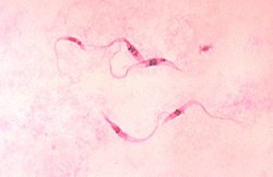
It can be used for histopathological diagnosis of the Plasmodium species that cause malaria[2] and some other spirochete and protozoan blood parasites.
It is also used to visualize the classic "safety pin" shape in Yersinia pestis.
This is particularly relevant for detection of Cytomegalovirus infection, where the classical finding would be an "owl-eye" viral inclusion.
[4] Giemsa stains the fungus Histoplasma, Chlamydia bacteria, and can be used to identify mast cells.