Human eye
The cornea is transparent and more curved and is linked to the larger posterior segment, composed of the vitreous, retina, choroid and the outer white shell called the sclera.
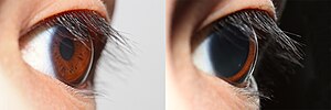
The iris is the pigmented circular structure concentrically surrounding the centre of the eye, the pupil, which appears to be black.
The size of the pupil, which controls the amount of light entering the eye, is adjusted by the iris' dilator and sphincter muscles.
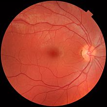
Photons of light falling on the light-sensitive cells of the retina (photoreceptor cones and rods) are converted into electrical signals that are transmitted to the brain by the optic nerve and interpreted as sight and vision.
The outermost layer, known as the fibrous tunic, is composed of the cornea and sclera, which provide shape to the eye and support the deeper structures.
The middle layer, known as the vascular tunic or uvea, consists of the choroid, ciliary body, pigmented epithelium and iris.
At the low end of the range is the absolute threshold of vision for a steady light across a wide field of view, about 10−6 cd/m2 (0.000001 candela per square meter).
[23][24] The upper end of the range is given in terms of normal visual performance as 108 cd/m2 (100,000,000 or one hundred million candelas per square meter).
[25] The eye includes a lens similar to lenses found in optical instruments such as cameras and the same physics principles can be applied.
Having two eyes allows the brain to determine the depth and distance of an object, called stereovision, and gives the sense of three-dimensionality to the vision.
This tracking is less accurate than the vestibulo-ocular reflex, as it requires the brain to process incoming visual information and supply feedback.
[35] Brown eyes result from a relatively high concentration of melanin in the stroma of the iris, which causes light of both shorter and longer wavelengths to be absorbed.
Although hazel eyes may contain specks of amber or gold, they usually tend to have many other colors, including green, brown and orange.
[41] Hazel eyes are due to a combination of Rayleigh scattering and a moderate amount of melanin in the iris' anterior border layer.
[35] A 2002 study found that the prevalence of blue eye color among the white population in the United States to be 33.8% for those born from 1936 through 1951.
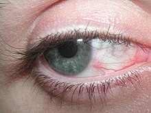
Related eye symptoms and signs of irritation are discomfort, dryness, excess tearing, itchiness, grating, foreign body sensation, ocular fatigue, pain, soreness, redness, swollen eyelids, and tiredness, etc.
[64][65] Eye irritation depends somewhat on destabilization of the outer-eye tear film, i.e. the formation of dry spots on the cornea, resulting in ocular discomfort.
[72][73] Other risk factors, such as chemical toxins/irritants (e.g. amines, formaldehyde, acetaldehyde, acrolein, N-Decane, VOCs, ozone, pesticides and preservatives, allergens, etc.)
[63] Nevertheless, if airborne particles alone should destabilize the tear film and cause eye irritation, their content of surface-active compounds must be high.
Dehydration, mental activities, work conditions, room temperature, relative humidity, and illumination all influence blink frequency.
This phenomenon indicates that perceived eye irritation is associated with an increase in blink frequency since the cornea and conjunctiva both have sensitive nerve endings that belong to the first trigeminal branch.
Field studies have found that the prevalence of objective eye signs is often significantly altered among office workers in comparisons with random samples of the general population.
[77][78][79][80] These research results might indicate that indoor air pollution has played an important role in causing eye irritation.
[93] A repeated measurement design was employed in the study of acute symptoms of eye and respiratory tract irritation resulting from occupational exposure to sodium borate dusts.
Results from multivariate logistic regression analysis suggest that current smokers tended to be less sensitive to the exposure to airborne sodium borate dust.
Therefore, for any detailed visually guided tasks on which performance varies with illumination, older persons require extra lighting.
Eye care professionals, including ophthalmologists and optometrists, are involved in the treatment and management of ocular and vision disorders.
Some disorders of the eyes for which corrective lenses are prescribed include myopia (near-sightedness), hyperopia (far-sightedness), astigmatism, and presbyopia (the loss of focusing range during aging).
[106] Lutein and zeaxanthin act as antioxidants that protect the retina and macula from oxidative damage from high-energy light waves.
[112] In the Renaissance, women used the juice of the berries of the belladonna plant in eyedrops to dilate the pupils and make the eyes appear more seductive.

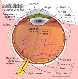


1. Lens , 2. Zonule of Zinn or Ciliary zonule , 3. Posterior chamber and 4. Anterior chamber with 5. Aqueous humour flow; 6. Pupil , 7. Corneosclera or Fibrous tunic with 8. Cornea , 9. Trabecular meshwork and Schlemm's canal . 10. Corneal limbus and 11. Sclera ; 12. Conjunctiva , 13. Uvea with 14. Iris , 15. Ciliary body (with a: pars plicata and b: pars plana ) and 16. Choroid ); 17. Ora serrata , 18. Vitreous humor with 19. Hyaloid canal/(old artery) , 20. Retina with 21. Macula or macula lutea , 22. Fovea and 23. Optic disc → blind spot ; 24. Visual axis (line of sight) . 25. Optical axis . 26. Optic nerve with 27. Dural sheath, 28. Tenon's capsule or bulbar sheath , 29. Tendon.
30. Anterior segment , 31. Posterior segment .
32. Ophthalmic artery , 33. Artery and central retinal vein → 36. Blood vessels of the retina; Ciliary arteries (34. Short posterior ones , 35. Long posterior ones and 37. Anterior ones ), 38. Lacrimal artery , 39. Ophthalmic vein , 40. Vorticose vein .
41. Ethmoid bone , 42. Medial rectus muscle , 43. Lateral rectus muscle , 44. Sphenoid bone .
