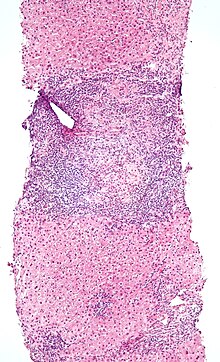
Intraepithelial lymphocyte
Upon encountering antigens, they immediately release cytokines and cause killing of infected target cells.
In both humans and mice IELs express higher levels of CD103, activation marker CD69, granzyme B and perforin cytolytic granules.
This molecule can bind MHC I, but, opposed to the function of CD8αβ, CD8αα reduces sensitivity of TCR towards antigens.
[8] More accurately, TL prevents proliferation of IELs, when there is co-occurrence of weak TCR stimulation.
IEL T cells acquire their activated memory phenotype post-thymically, in response to antigens encountered in the periphery.
[11] Their role in immune system is crucial because IELs provide a first line of defense at this extensive barrier with the outside world.
IELs mediate antigen-specific delayed-type hypersensitivity (DTH) responses, exhibit virus-specific CTL function, to express natural killer (NK)-like activity and produce a local graft-versus-host reaction (GVHR) when transferred to semiallogeneic hosts.
Also termed conventional IELs, express TCRαβ together with CD4 or CD8αβ and are derived from antigen-experienced T cells that home to intraepithelial space.
In mice, up to 50% of these IELs can express CD8αα homodimer, which they acquire in the intestinal epithelium after external stimuli such as TGF-β, IFN-γ, IL-27 and retinoic acid.
[3][17] These IELs emerge from peripherally activated conventional CD8+ T-cells and home to the intestinal epithelium, where they function as effector or memory cells.
They continuously express integrin β7, granzyme B, CD103 and CD69 and produce lower amounts of TNF-α and IFN-γ as opposed to the conventional CD8+ T-cells.
[20] Another transcription factor responsible for DP IELs induction is the Aryl hydrocarbon receptor (AhR).
The function of CD4CD8aa IELs is due to their CD8 phenotype and granzyme B expression to prevent pathogens from invading and to maintain integrity of the intestinal epithelial barrier.
Their CD4 phenotype is responsible for IL-10 and TGF-β secretion that prevents Th1-induced inflammation in the intestine, therefore their role can be complementary to T regulatory cells.
[3] TCRγδ+ IELs develop outside of thymus and their maintenance and function in the intestinal epithelium is influenced by a cross-talk with enterocytes.
The mechanism is not clear, but TCRγδ+ IELs have cytotoxic properties and can produce cytokines TGF-β, TNF-α, IFN-γ, IL-13 and IL-10 and antimicrobial peptides, all of which can contribute to the diverse functions.
[3] Similar functions have been found in the context of colitis, where these cells seem to have pathogenic role at the beginning, whereas later they protect the epithelium against tissue damage.
They have cytotoxic and phagocytic properties, express MHC II and thereby can present antigens to conventional CD4+ T-cells.
