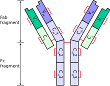
Immunoglobulin light chain
The immunoglobulin light chain genes in tetrapods can be classified into three distinct groups: kappa (κ), lambda (λ), and sigma (σ).
[5] Sharks also possess, as part of their adaptive immune systems, a functional heavy-chain homodimeric antibody-like molecule referred to as IgNAR (immunoglobulin new antigen receptor).
Using immunohistochemistry, it is possible to determine the relative abundance of B-cells expressing kappa and lambda light chains.
If, however, one type of light chain is significantly more common than the other, the cells are likely all derived from a small clonal population, which may indicate a malignant condition, such as B-cell lymphoma.
[9] Free immunoglobulin light chains secreted by neoplastic plasma cells, such as in multiple myeloma, can be called Bence Jones protein when detected in the urine, although there is a trend to refer to these as urinary free light chains.
It is important to note that, in contrast to increased levels in lymphoma patients, these Ig light chains are polyclonal.
[10] Activation of mast cells results in the release of various pro-inflammatory mediators which are believed to contribute to the development of the inflammatory disease.
Recent studies have shown that Ig light chains not only activate mast cells but also dorsal root ganglia[11] and neutrophils,[12] expanding their possible role as mediators in inflammatory disease.

![[1]](https://web.archive.org/web/20070419154840/http://www.emc.maricopa.edu/faculty/farabee/BIOBK/ANTIBODY.gif)