Maximum intensity projection
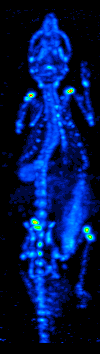
MIP imaging is also used routinely by physicians in interpreting Positron Emission Tomography (PET) or Magnetic Resonance Angiography studies.
To improve the sense of 3D, animations are usually rendered of several MIP frames in which the viewpoint is slightly changed from one to the other, thus creating the illusion of rotation.
However, since the projection is orthographic the viewer cannot distinguish between left or right, front or back and even if the object is rotating clockwise or anti-clockwise.
Because - in general - we can terminate the ray earlier this technique is faster and also can give better results in some settings as it approximates occlusion.
MIP Display was invented for use in Nuclear Medicine by Jerold Wallis, MD, in 1988 at Washington University in St. Louis, and subsequently published in IEEE Transactions on Medical Imaging.

- Average intensity projection
- Maximum intensity projection
- Thin slice ( median plane )
- Volume rendering by high and low threshold for radiodensity .