Metallography
Different materials with similar properties (hardness and ductility) will respond alike and thus require the same consumables during preparation.
In the past, phenolic thermosetting resins have been used, but modern epoxy is becoming more popular because reduced shrinkage during curing results in a better mount with superior edge retention.
Typically, a specimen is polished with a slurry of alumina, silica, or diamond on a napless cloth to produce a scratch-free mirror finish, free from smear, drag, or pull-outs and with minimal deformation remaining from the preparation process.
Non-destructive surface analysis techniques can involve applying a thin film or varnish that can be peeled off after drying and examined under a microscope.
Thus, the analysis can determine if the more expensive, more time-consuming examination techniques using the SEM or the TEM are required and where on the specimen the work should be concentrated.
Special methods are used at magnifications below 50X, which can be very helpful when examining the microstructure of cast specimens where greater spatial coverage in the field of view may be required to observe features such as dendrites.
Image contrast depends upon the quality of the optics, coatings on the lenses, and reduction of flare and glare; but, it also requires proper specimen preparation and good etching techniques.
Most LOM observations are conducted using bright-field (BF) illumination, where the image of any flat feature perpendicular to the incident light path is bright, or appears to be white.
Dark-field microscopy (DF), is an alternative method of observation that provides high-contrast images and actually greater resolution than bright-field.
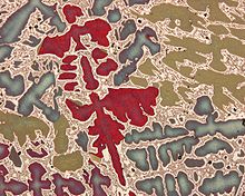
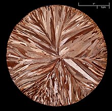
If the specimen is prepared with minimal damage to the surface, the structure can be seen vividly in cross-polarized light (the optic axis of the polarizer and analyzer are 90 degrees to each other, i.e., crossed).
Another useful imaging mode is differential interference contrast (DIC), which is usually obtained with a system designed by the Polish physicist Georges Nomarski.
DIC has largely replaced the older oblique illumination (OI) technique, which was available on reflected light microscopes prior to about 1975.
In OI, the vertical illuminator is offset from perpendicular, producing shading effects that reveal height differences.
OI can be created on any microscope by placing a piece of paper under one corner of the mount so that the plane-of-polish is no longer perpendicular to the optical axis.
The ability to detect low-atomic number elements, such as carbon, oxygen, and nitrogen, depends upon the nature of the detector used.
But, quantification of these elements by EDS is difficult and their minimum detectable limits are higher than when a wavelength-dispersive spectrometer (WDS) is used.
If a particular phase can be chemically extracted from a bulk specimen, it can be identified using XRD based on the crystal structure and lattice dimensions.
Stereology is the field of taking 0-, 1- or 2-dimensional measurements on the two-dimensional sectioning plane and estimating the amount, size, shape or distribution of the microstructure in three dimensions.
These measurements may be made using manual procedures with the aid of templates overlaying the microstructure, or with automated image analyzers.
A stereological method for characterizing discrete second-phase particles, such as nonmetallic inclusions, carbides, graphite, etc., is presented in ASTM E 1245.