Microfluorimetry
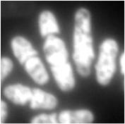
This technique allows for karyotyping at higher speeds than with previous methods and was shown to be accurate using Chinese hamster chromosomes.
The markers used for measurement in flow microfluorimetry are made up of fluorescent antigens or DNA binding agents.
[4] For example, microfluorimetry is used in neurons to compare the effects of neurotoxins on both calcium ion concentration and mitochondrial membrane potential in individual cells.
[5] Microfluorimetry can also be used as a method to distinguish different microorganisms from one another by analyzing and comparing the DNA content of each cell.
[7] Another use of microfluorometry is flow cytometry which uses the emission of fluorochrome molecules and usually a laser as a light source to create data from particles and cells.
[9] For example, E. coli bacteriophages lambda and T4 were able to be separated by flow cytometry which allowed for genomic analysis which was previously difficult.