Microsporum canis
Microsporum canis is a pathogenic, asexual fungus in the phylum Ascomycota that infects the upper, dead layers of skin on domesticated cats, and occasionally dogs and humans.
[3] Microsporum canis reproduces by means of two conidial forms, large, spindle-shaped, multicelled macroconidia and small, single-celled microconidia.
Microsporum canis exemplifies a common situation in ascomycetous fungi in which, over time, one mating type strain has undergone habitat divergence from the other and established a self-sustaining reproductive population that consists only of the asexual form.
[3] It is hypothesized that asexual lineage of Microsporum canis evolved as a result of host-specific interactions, changes in ecological niche, as well as, geographic isolation of + and – mating types of Arthroderma otae, hence making it difficult to sustain sexual reproduction.
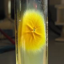
[4] Some strains of M. canis fail to produce yellow pigment altogether, exhibit abnormally slow colony growth and form undeveloped macroconidia.
[4] Microsporum canis reproduces asexually by forming macroconidia that are asymmetrical, spindle-shaped and have cell walls that are thick and coarsely roughened.
[3][4] Microsporum canis produces infections of scalp and body sites, creating highly inflammatory lesions associated with hair loss.
[3] Despite the frequent use of Wood's lamp in the clinical evaluation of ringworm infections, diagnosis of M. canis requires the performance of additional tests given the potential for false positives.
[5][12] Genetic analyses can be useful to establish the identity of atypical strains of M. canis; however the highly characteristic appearance of this species generally obviates the need for this more sophisticated method.
[4] This colonization of the hair shaft causes it to become unsheathed, resulting in characteristic round or oval non-inflammatory lesions the develop on the scalp.
[4][9] Infection triggers an acute leukocytic reaction in subcutaneous tissues, which gradually becomes highly inflammatory and leads to hair loss, in the case of tinea capitis.
[10] Microsporum canis infections can be easily managed by topical antifungal agents; however severe cases may necessitate systemic therapy with griseofulvin, itraconazole or terbinafine.