Myofibril
These proteins are organized into thick, thin, and elastic myofilaments, which repeat along the length of the myofibril in sections or units of contraction called sarcomeres.
Exposed muscle cells at certain angles, such as in meat cuts, can show structural coloration or iridescence due to this periodic alignment of the fibrils and sarcomeres.
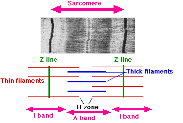
[5] The names of the various sub-regions of the sarcomere are based on their relatively lighter or darker appearance when viewed through the light microscope.
Each sarcomere is delimited by two very dark colored bands called Z-discs or Z-lines (from the German zwischen meaning between).
The I bands appear lighter because these regions of the sarcomere mainly contain the thin actin filaments, whose smaller diameter allows the passage of light between them.
A stands for anisotropic and I for isotropic, referring to the optical properties of living muscle as demonstrated with polarized light microscopy.
Also within the A band is a relatively brighter central region called the H-zone (from the German helle, meaning bright) in which there is no actin/myosin overlap when the muscle is in a relaxed state.
Finally, the H-zone is bisected by a dark central line called the M-line (from the German mittel meaning middle).
Along the long axis of the muscle cells in subsarcolemmal locations, free myofilaments become aligned and aggregate into hexagonally packed arrays.
When the muscle fibre is relaxed (before contraction), the myosin head has ADP and phosphate bound to it.