Myostatin
Human myostatin consists of two identical subunits, each consisting of 109 (NCBI database claims human myostatin is 375 residues long) amino acid residues [note the full length gene encodes a 375AA prepro-protein which is proteolytically processed to its shorter active form].
The protein is inactive until a protease cleaves the NH2-terminal, or "pro-domain" portion of the molecule, resulting in the active COOH-terminal dimer.
This coreceptor then initiates a cell signaling cascade in the muscle that includes the activation of transcription factors in the SMAD family—SMAD2 and SMAD3.
[21] Mutations in myostatin do more than just affect the amount of muscle mass an organism can produce; they also have variable effects on other phenotypes for different species.
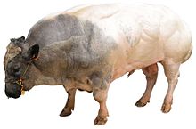
[21]For example, a Belgian Blue bovine with a mutation that inhibits myostatin production will exhibit a dramatic increase in muscle mass but will also lead to dystocia.
[25] However, the animal breeds developed as homozygous for myostatin deficiency have reproduction issues due to their unusually heavy and bulky offspring, and require special care and a more expensive diet to achieve a superior yield.
Animals with a homozygous deletion have an unusual body shape, with a broader head, pronounced overbite, shorter legs, and thicker tails, and are called "bully whippets" by the breeding community.
A South Korean-Chinese team has engineered "double muscle" pigs, as with cattle, aiming for cheaper breeds for the meat market.
People with a mutation in both copies of the MSTN gene in each cell (homozygotes) have significantly increased muscle mass and strength.
People with a mutation in one copy of the MSTN gene in each cell (heterozygotes) have increased muscle bulk, but to a lesser degree.
[citation needed] In 2004, a German boy was diagnosed with a mutation in both copies of the myostatin-producing gene, making him considerably stronger than his peers.
The mice were sent to the International Space Station and could largely maintain their muscle weights – about twice those of wild type due to genetic engineering for targeted deletion of the myostatin gene – under microgravity.
[46] It remains unclear as to whether long-term treatment of muscular dystrophy with myostatin inhibitors is beneficial, as the depletion of muscle stem cells could worsen the disease later on.
[58][56] Knockdown of myostatin has been shown to reduce formation of osteoclasts (multinucleated cells responsible for the breakdown of bone tissue) in mice modeling rheumatoid arthritis.
[58] Rheumatoid arthritis is an autoimmune disorder that, among other effects, leads to the degradation of the bone tissue in affected joints.
One study[58] by Berno Dankbar et al., 2015 found that myostatin deficiency leads to a notable reduction in inflammation around a fracture site.
[58][56] An association between osteoporosis, another disease characterized by the degradation of bony tissue, and sarcopenia, the age-related degeneration of muscle mass and quality have also been found.
[61] Pathological cardiac stress promotes N-terminal cleavage by furin convertase to create a biologically active C-terminal fragment.
[61] The latter produces a heteromeric complex with SMAD4, inducing myostatin translocation into the cardiomyocyte nucleus to modulate transcription factor activity.
[61][63] Physiologically, minimal amounts of cardiac myostatin are secreted from the myocardium into serum, having a limited effect on muscle growth.
[62][63] It has been hypothesized that hypertrophy of the heart induces an increase in myostatin as a negative feedback mechanism in an attempt to limit further myocyte growth.
[67][68] This process includes mitogen-activated protein kinases and binding of the MEF2 transcription factor within the promoter region of the myostatin gene.
[61][62][69] Systemic inhibition of cardiac myostatin with the JA-16 antibody maintains overall muscle weight in experimental models with pre-existing heart failure.
[70] A reduction in cardiac myostatin induces eccentric hypertrophy of the heart, and increases its sensitivity to beta-adrenergic stimuli by enhancing Ca2+ release from the SR during EC coupling.