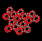
Nanosponges
[1] Their small size allows them to move quickly through substances, like water or blood, efficiently finding and attacking unwanted matter.
Nanosponges are superior to microsponges in application as the smaller size allows less disruption into the system in which it is implemented therefore imposing less risk of failed or detrimental effects.
This useful structure allows them to act as drug carriers in the body, as long as the compounds to be delivered have compatible geometry and polarity with the cavity.
Researchers have found promising results in using these nanosponges in drug delivery, relieving inflammation, and repairing damaged tissue.
Other functions of these nanorobots are the ability to neutralize cytolytic activity regardless of the molecular structure, enhancing mass transport, and they may also be able to fight auto-immune diseases.
Engineers at Cornell University have created a 20 nanometer long particle that can self assemble in water so that its orientation allows for a hydrophilic exterior and hydrophobic interior.
The Cornell researchers injected these nanoparticles into the bottom of a steel column filled with sand contaminated by phenanthrene and a polycyclic aromatic hydrocarbon (PAH), components typically found in tar.
These effects include allergic reactions, insomnia, vision problems, and can be as extreme as to cause mental disability, dementia, and kidney disease.
Using nanosponges for this results in a higher efficiency and lower cost than alternative cleaning methods like ion exchange resins, activated carbon, or other biological agents.
Porous materials produced from renewable and low cost sources, like cellulose, chitin, or starch, are one of the most promising classes of absorbents in terms of effectiveness.
[1] Although the research of these citric acid nanosponges is still undergoing revision and development, they show promise for being a sustainable way to clean metal deposits from the ecosystem.
[8] Major concerns regarding recently developed chemical entities include pharmacokinetic issues, poor solubility in water, and low bioavailability.
Nanosponges can conquer these problems as their porous structure allows them the unique capability to entrap both hydrophilic and hydrophobic drugs and release them in a highly predictable manor.
These small sponges travel throughout the body until they reach the targeted site where they bind to the surface and perform controlled drug release.
These small sponges travel throughout the body until they reach the targeted site where they bind to the surface and perform controlled drug release.
The study was conducted using a nanosponge (polymeric core) wrapped in a natural red blood cell membrane bilayer so that the bacteria or venom will attack it.
Due to enhanced responsiveness of conductance to surface effects, various forms of metal oxides that have been nanostructured have been synthesized and their sensing properties studied.
The best SERS enhancement is achieved by having strong localized plasmons that fall within the wavelength of the Raman laser excitation, which is why gold and silver are often used.
Fluorescence arises from photoexcitation in these quantum dots and is easily tuned to the visible or near infrared region of the spectrum, by choice of semiconductor material and particle size, making quantum-useful fluorophores.
They lend themselves nicely to multichannel fluorophore systems, with single excitation wavelength causing emission from many species of many different colors.
[8] A lot of research is only in its primary stages as implementing these solution to the human body poses many risks for which these applications of nanosponges are not yet developed enough.
[15] After head injury, mice were left to be for two to three hours and subsequently injected with biodegradable nanoparticles made from an unspecified but FDA approved polymer which is commonly used in some dissolving sutures.
[3] Because the elimination of these particles can happen so fast, researchers were able to inject mice once more two to three days later to combat inflammation that might come back slowly after injury.
[3] Mice's vision cells performed better in response to light and were able to better walk across a ladder after recovering showing improvement in behavior and motor function.
These have been tested in a mouse Escherichia coli bacteremia model, where the nanoparticles were able to significantly increase the survival of the mice by decreasing the proinflammatory cytokine levels and preventing the bacteria from disseminating.
[19][20] Mn3O4@nanoerythrocyte-T7 (MNET) nanosponges can regulate oxygen and scavenge free radicals in the event of an ischemic stroke, which is a global leading cause of death and disability.
These engineered nanosponges can help attenuate hypoxia after a stroke by mimicking red blood cells and increasing the amount of oxygen in the infarct area.
This allows for the extension of the survival time of neurocytes, a crucial part of treating an ischemic stroke because their normal functions must be maintained.
[22] In a study on middle cerebral artery occlusion (MCAO) rats, those treated with MNET experienced a significant attenuation of neurological damage.
For example, Dr. Zhang of the University of California, San Diego suggests that for rheumatoid arthritis, this could elicit an immune response, therefore, not fighting the disease but driving it.