Neural plate
In embryology, the neural plate is a key developmental structure that serves as the basis for the nervous system.
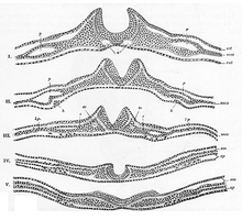
[citation needed] Stretched over the notochord, the ectodermal cells on the dorsal portion of the embryo are ultimately the ones that form the neural plate.
These regions furrow and change shape in the same way as MHP cells do before connecting together to form the neural tube.
[7] Critical to the proper folding and function of the neural plate is N-cadherin, a type of cadherin protein associated with the nervous system.
Axial mesoderm cells under the ectoderm secrete inhibitory signals called chordin, noggin and follistatin.
[9] These fluctuations in mRNA and protein expression allude to how they play a role in differentiation of neural plate cells.
Low pSMAD 1, 5, 8 levels allow a greater mobility at the median hinge point than in lateral neural plate cells.
In chickens, neural tube closure begins at the future midbrain region and it closes in both directions.
The newt neural plate doubles in length, decreases in apical width, and increases in thickness.
[5] Research on the neural plate began in earnest by looking into the determination of the ectoderm and its commitment to the neuronal path.
The use of such techniques vary with the stage of development and overall research goals, but include such methods as cell labeling and grafting.
This technique is useful as it reveals specific areas of gene expression in a tissue as well as throughout an entire embryo through whole-mount in situ hybridization.
Similar to the process of in situ hybridization, immunofluorescence (IF) also allows for the determination of particular cell element's roles in development.
In contrast to in situ hybridization however, immunofluorescence uses a fluorophore attached to an antibody with biomolecule target, such as proteins, rather than DNA and RNA sequences.
In the study of embryogenesis immunofluorescence may be used for purposes similar to hybridization, for the tracking of proteins that are involved in the development of the embryo and their specific time and place of production and use.
[13] Current research has expanded on the immunofluorescence technique to combined it with the methods of in situ hybridization, either fluorescent or radioactive.
Grafting at specific stages of neurulation has advanced research on the signaling necessary for the proper development of the neural plate and other structures.
The grafting of the ectoderm and neural structures is very specialized and delicate procedure, requiring the removal and marking of a desired group of cells, followed by their transplantation, for example, into a new area of the embryo.