Neurulation
The process begins when the notochord induces the formation of the central nervous system (CNS) by signaling the ectoderm germ layer above it to form the thick and flat neural plate.
[2] Computer simulations found that cell wedging and differential proliferation are sufficient for mammalian neurulation.
[3] Different portions of the neural tube form by two different processes, called primary and secondary neurulation, in different species.
[5] The first experiments proving induction were attributed by Viktor Hamburger[6] to independent discoveries of both Hans Spemann of Germany in 1901[7] and Warren Lewis of the USA in 1904.
[12] Items as diverse as low pH, cyclic AMP, even floor dust could act as inducers leading to considerable consternation.
[22][23] There has long been a general reluctance in the field to consider the possibility that primary neural induction might be initiated by mechanical effects.
As neurulation proceeds after induction, the cells of the neural plate become high-columnar and can be identified through microscopy as different from the surrounding presumptive epithelial ectoderm (epiblastic endoderm in amniotes).
This pyramid shape is achieved through tubulin and actin in the apical portion of the cell which constricts as they move.
As a result of the cellular shape changes, the neural plate forms the medial hinge point (MHP).
Instead, it begins approximately at the level of the fourth somite at Carnegie stage 9 (around embryonic day 20 in humans).
[28] Failure of the cranial (superior) and caudal (inferior) neuropore closure results in conditions called anencephaly and spina bifida, respectively.
Additionally, failure of the neural tube to close throughout the length of the body results in a condition called rachischisis.
For example, retained medullary cord occurs due to a partial or complete arrest of secondary neurulation that creates a non-functional portion on the vestigial end.
[36] The anterior portion of the neural tube forms the three main parts of the brain: the forebrain (prosencephalon), midbrain (mesencephalon), and the hindbrain (rhombencephalon).
The hindbrain, which is the evolutionarily most ancient part of the chordate brain, also divides into different segments called rhombomeres.
The rhombomeres generate many of the most essential neural circuits needed for life, including those that control respiration and heart rate, and produce most of the cranial nerves.
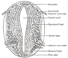
The early neural tube is primarily composed of the germinal neuroepithelium, later called the ventricular zone, which contains primary neural stem cells called radial glial cells and serves as the main source of neurons produced during brain development through the process of neurogenesis.
[39][40] Paraxial mesoderm surrounding the notochord at the sides will develop into the somites (future muscles, bones, and contributes to the formation of limbs of the vertebrate ).
[42] Failure of the rostral end of the neural tube to close results in anencephaly, or lack of brain development, and is most often fatal.