Neuroanatomy
For example, much of what neuroscientists have learned comes from observing how damage or "lesions" to specific brain areas affects behavior or other neural functions.
The first known written record of a study of the anatomy of the human brain is an ancient Egyptian document, the Edwin Smith Papyrus.
Herophilus and Erasistratus of Alexandria were perhaps the most influential with their studies involving dissecting human brains, affirming the distinction between the cerebrum and the cerebellum, and identifying the ventricles and the dura mater.
[4][5] The Greek physician and philosopher Galen, likewise, argued strongly for the brain as the organ responsible for sensation and voluntary motion, as evidenced by his research on the neuroanatomy of oxen, Barbary apes, and other animals.
[3][6] The cultural taboo on human dissection continued for several hundred years afterward, which brought no major progress in the understanding of the anatomy of the brain or of the nervous system.
However, Pope Sixtus IV effectively revitalized the study of neuroanatomy by altering the papal policy and allowing human dissection.
This resulted in a flush of new activity by artists and scientists of the Renaissance,[7] such as Mondino de Luzzi, Berengario da Carpi, and Jacques Dubois, and culminating in the work of Andreas Vesalius.
[8][9] In 1664, Thomas Willis, a physician and professor at Oxford University, coined the term neurology when he published his text Cerebri Anatome which is considered the foundation of modern neuroanatomy.
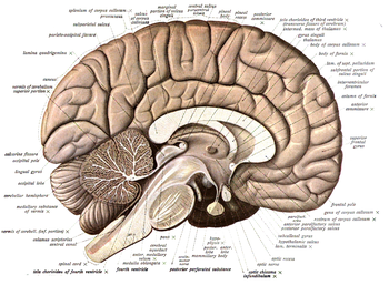
At the organ level, the nervous system is composed of brain regions, such as the hippocampus in mammals or the mushroom bodies of the fruit fly.
Again in this case, the situation is different for swimming, creeping or quadrupedal (prone) animals than for Man, or other erect species, due to the changed position of the axis.
This so-called silver chromate impregnation procedure stains entirely or partially the cell bodies and neurites of some neurons -dendrites, axon- in brown and black, allowing researchers to trace their paths up to their thinnest terminal branches in a slice of nervous tissue, thanks to the transparency consequent to the lack of staining in the majority of surrounding cells.
Modernly, Golgi-impregnated material has been adapted for electron-microscopic visualization of the unstained elements surrounding the stained processes and cell bodies, thus adding further resolutive power.
This immensely increases the capacity of researchers to distinguish between different cell types (such as neurons and glia) in various regions of the nervous system.
By expressing variable amounts of red, green, and blue fluorescent proteins in the brain, the so-called "brainbow" mutant mouse allows the combinatorial visualization of many different colors in neurons.
Optogenetics uses transgenic constitutive and site-specific expression (normally in mice) of blocked markers that can be activated selectively by illumination with a light beam.
Magnetic resonance imaging has been used extensively to investigate brain structure and function non-invasively in healthy human subjects.
[18] Axonal transport methods use a variety of dyes (horseradish peroxidase variants, fluorescent or radioactive markers, lectins, dextrans) that are more or less avidly absorbed by neurons or their processes.
[19] Circuit reconstruction from data produced by this high-throughput method is challenging, and the Citizen science game EyeWire has been developed to aid research in that area.
Is a field that utilizes various imaging modalities and computational techniques to model and quantify the spatiotemporal dynamics of neuroanatomical structures in both normal and clinical populations.
The mouse is used because, as a mammal, its brain is more similar in structure to our own (e.g., it has a six-layered cortex, yet its genes can be easily modified and its reproductive cycle is relatively fast).
In spite of the large evolutionary distance between insects and mammals, many basic aspects of Drosophila neurogenetics have turned out to be relevant to humans.
For instance, the first biological clock genes were identified by examining Drosophila mutants that showed disrupted daily activity cycles.