Paracoccidioidomycosis
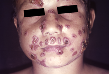
[14] Skin and mucous membrane lesions are often present,[17] and bone involvement may occur in severe cases.
[14] Upper respiratory tract mucosal lesions may be present, as well as increased mucus production and coughing up blood.
[15] Paracoccidioidomycosis is caused by two species of fungi that can exist as a mold or yeast depending on temperature, Paracoccidioides brasiliensis and P.
[19] In protected soil environments, near water sources, that are disturbed either naturally or by human activity, P. brasiliensis has been epidemiologically observed (although not isolated).
[13] In the natural environment, the fungi are found as filamentous structures, and they develop infectious spores known as conidia.
[15] After inhalation into the alveoli, there is rapid multiplication of the organism in the lung tissue, sometimes spreading via the venous and lymphatic systems.
[15] The type of immune response determines the clinical manifestation of the infection, with children and HIV co-infected individuals most commonly developing the acute/subacute disseminated disease.
In these individuals, granulomas do not form, and the affected person develops Th2 and Th9 responses, resulting in activation of B lymphocytes, high levels of circulating antibodies, eosinophilia, and hypergammaglobulinemia.
[15] The commonest, chronic form, is almost certainly a reactivation of the disease,[15] and may develop into progressive scarring of the lungs (pulmonary fibrosis).
[23] More than 90% of cases can be diagnosed with a direct histological examination of tissue, such as sputum, bronchial lavage fluid, exudates, and biopsies.
[14] Complement fixation allows for a measure of severity of cases by quantifying the antibody level, and is thus useful for monitoring treatment response.
[14] The disease can appear similar to tuberculosis, leukaemia, and lymphoma[4] Both P. brasiliensis and P. lutzii are in-vitro susceptible to most antifungal agents, unlike other systemic fungal infections.
Mild and moderate forms are treated with itraconazole for 9 to 18 months, as this is more effective, has a shorter treatment duration, and is more tolerated.
[25] Paracoccidioidomycosis is endemic in rural areas of Latin America, from southern Mexico to Argentina, and is also found in Brazil, Colombia, Venezuela, Ecuador, and Paraguay.
[21] Rising cases have been linked to agriculturalization and deforestation in Brazil, urbanisation to peripheral city areas with poor infrastructure, as well as increased soil and air humidity.
[21] Lutz-Splendore-de Almeida disease[3] is named for the physicians Adolfo Lutz,[27] Alfonso Splendore [pt] (1871–1953), an Italo-Brazilian parasitologist[28] and Floriano Paulo de Almeida (1898–1977), a Brazilian pathologist specializing in Pathologic Mycology (Study of Infectious Fungi),[29][30] who first characterized the disease in Brazil in the early 20th century.