Phycomycosis
Phycomycosis is an uncommon condition affecting the gastrointestinal tract and skin, most commonly found in dogs and horses.
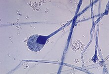
The condition is caused by various molds (a type of fungi), with individual forms including pythiosis, zygomycosis, and lagenidiosis.
Zygomycosis can be caused by two types of zygomycetes: Entomophthorales (e.g., Basidiobolus and Conidiobolus) and Mucorales (e.g., Mucor, Mortierella, Absidia, Rhizopus, Rhizomucor, and Saksenaea).
Pythiosis is caused by Pythium insidiosum and occurs most commonly in dogs and horses, but is also found in cats, cattle, and humans.
Pythiosis occurs in areas with mild winters due to the organism surviving in standing water that does not reach freezing temperatures.
[2] The disease grows slowly in the stomach and small intestine, eventually forming large lumps of granulation tissue.
In horses, subcutaneous pythiosis is the most common form and infection occurs through a wound while standing in water containing the pathogen.
[9] Entomophthorales is found in soil and decaying plant matter, and specifically, Basidiobolus can be contracted from insects and the feces of reptiles or amphibians.
Lagenidiosis causes progressive skin and subcutaneous lesions in the legs, groin, trunk, and near the tail.
Spread of the disease to distant lymph nodes, large blood vessels, and the lungs may occur.
[10] Diagnosis is through biopsy or culture, although an enzyme-linked immunosorbent assay (ELISA) test has been developed for Pythium insidiosum in animals.
Antifungal drugs show only limited effect on the disease, but itraconazole and terbinafine hydrochloride are often used for 2 to 3 months following surgery.