Pupil
[citation needed] The size of the pupil is controlled by the iris, and varies depending on many factors, the most significant being the amount of light in the environment.
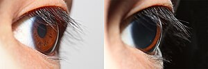
[7] When bright light is shone on the eye, light-sensitive cells in the retina, including rod and cone photoreceptors and melanopsin ganglion cells, will send signals to the oculomotor nerve, specifically the parasympathetic part coming from the Edinger-Westphal nucleus, which terminates on the circular iris sphincter muscle.
[citation needed] If the drug pilocarpine is administered, the pupils will constrict and accommodation is increased due to the parasympathetic action on the circular muscle fibers, conversely, atropine will cause paralysis of accommodation (cycloplegia) and dilation of the pupil.
[8] Other drugs, such as atropine, LSD, MDMA, mescaline, psilocybin mushrooms, cocaine and amphetamines may cause pupil dilation.
In pupillary constriction induced by pilocarpine, not only is the sphincter nerve supply activated but that of the dilator is inhibited.
This condition is typified by chronically widened pupils due to the decreased ability of the optic nerves to respond to light.
It is necessary for these people to be especially careful when driving at night due to their inability to see objects in their full perspective.
In addition to dilation and contraction caused by light and darkness, it has been shown that solving simple multiplication problems affects the size of the pupil.
[13] There is also evidence that pupil size is related to the extent of positive or negative emotional arousal experienced by a person.
[15] Some humans are able to exert direct control over their iris muscles, giving them the ability to manipulate the size of their pupils (i.e. dilating and constricting them) on command, without any changes in lighting condition or eye accommodation state.
Some have slits or ovals which may be oriented vertically, as in crocodiles, vipers, cats and foxes, or horizontally as in some rays, flying frogs, mongooses and artiodactyls such as elk, red deer, reindeer and hippopotamus, as well as the domestic horse.
Goats, sheep, toads and octopus pupils tend to be horizontal and rectangular with rounded corners.
[21] An alternative explanation is that a partially constricted circular pupil shades the peripheral zones of the lens which would lead to poorly focused images at relevant wavelengths.
[3] It has also been suggested that in ambush predators such as some snakes, vertical slit pupils may aid in camouflage, breaking up the circular outline of the eye.
[22] In a study of Australian snakes, pupil shapes correlated both with diel activity times and with foraging behavior.
It has been suggested that there may be a similar link between foraging behaviour and pupil shape amongst the felidae and canidae discussed above.
[22] A 2015 study[23] confirmed the hypothesis that elongated pupils have increased dynamic range, and furthered the correlations with diel activity.
[25] In the Old Babylonian period (c. 1800-1600 BC) in ancient Mesopotamia, the expression "protective spirit of the eye" is attested, perhaps arising from the same phenomenon.