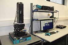
Raman microscope
In direct imaging, the whole field of view is examined for scattering over a small range of wavenumbers (Raman shifts).
[4][5][6] Sophisticated signal- and image-processing techniques can be used to ignore the presence of water, culture media, buffers, and other interference.
[7] Because a Raman microscope is a diffraction-limited system, its spatial resolution depends on the wavelength of light and the numerical aperture of the focusing element.
This means that when operated in the visible to near-infrared range, a Raman microscope can achieve lateral resolutions of approx.
[8][9][10] Since the objective lenses of microscopes focus the laser beam down to the micrometer range, the resulting photon flux is much higher than achieved in conventional Raman setups.
Raman microscopy of inorganic specimens, such as rocks, ceramics and polymers,[13] can use a broader range of excitation wavelengths.
A related technique, tip-enhanced Raman spectroscopy, can produce high-resolution hyperspectral images of single molecules[14] and DNA.
Consequently, in vivo time- and space-resolved Raman spectroscopy is suitable to examine proteins, cells and organs.
Combining stable isotopic probing (SIP) experiments with confocal Raman microspectroscopy has permitted determination of assimilation rates of 13C and 15N-substrates as well as D2O by individual bacterial cells.