SEEC microscopy
Surface-enhanced ellipsometric contrast microscopy (SEEC) uses an upright or inverted optical microscope in a crossed polarization configuration and specific supporting plates called surfs on which the sample is deposited for observation.
SEEC relies on precise control of the reflection properties of polarized light on a surface, improving the axial sensitivity of an optical microscope by two orders of magnitude without reducing its lateral resolution.
A 2006 study on polarized light coherence led to the development of new supports (the surfs) having contrast amplification properties for standard optical microscopy in cross-polarizer mode.
[2] Made of optical layers on an opaque or transparent substrate, these supports do not modify the light polarization after reflection even if the numerical aperture of the incident source is significant.
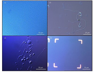
An illustration of the contrast enhancement is in the Figure for optical microscopy between cross polarizers of a Langmuir-Blodgett structure on a silicon wafer and on a surf.