Crinoid
This is surrounded by feeding arms, and is linked to a U-shaped gut, with the anus being located on the oral disc near the mouth.
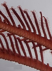
Although the basic echinoderm pattern of fivefold symmetry can be recognised, in most crinoids the five arms are subdivided into ten or more.
Some thick limestone beds dating to the mid-Paleozoic era to Jurassic period are almost entirely made up of disarticulated crinoid fragments.
[14] The numerous calcareous plates make up the bulk of the crinoid, with only a small percentage of soft tissue.
These ossicles fossilise well and there are beds of limestone dating from the Lower Carboniferous around Clitheroe, England, formed almost exclusively from a diverse fauna of crinoid fossils.
[15] The stem of sea lilies is composed of a column of highly porous ossicles which are connected by ligamentary tissue.
[5] Crinoids are passive suspension feeders, filtering plankton and small particles of detritus from the sea water flowing past them with their feather-like arms.
Mobile crinoids move to perch on rocks, coral heads or other eminences to maximise their feeding opportunities.
There is no true stomach, so the oesophagus connects directly to the intestine, which runs in a single loop right around the inside of the calyx.
[5] Specimens of the sea urchin Calocidaris micans found in the vicinity of the crinoid Endoxocrinus parrae, have been shown to contain large quantities of stem portions in their guts.
[20] Like other echinoderms, crinoids possess a water vascular system that maintains hydraulic pressure in the tube feet.
The action of cilia cause there to be a slow flow of fluid (1mm per second) in these canals, outward in the oral branches and inward in the aboral ones, and this is the main means of transport of nutrients and waste products.
There is no heart and separate circulatory system but at the base of the disc there is a large blood vessel known as the axial organ, containing some slender blind-ended tubes of unknown function, which extends into the stalk.
Oxygen is absorbed primarily through the tube feet, which are the most thin-walled parts of the body, with further gas exchange taking place over the large surface area of the arms.
The third portion of the nervous system lies aborally, and is responsible for the flexing and movement actions of the arms, pinnules and cirri.
The bilaterally symmetrical larva is barrel-shaped with rings of cilia running round the body, and a tuft of sensory hairs at the upper pole.
[21] The larva's free-swimming period lasts for only a few days before it settles on the bottom and attaches itself to the underlying surface using an adhesive gland on its underside.
Arms torn off by predators or damaged by adverse environmental conditions can regrow, and even the visceral mass can regenerate over the course of a few weeks.
Such a movement may be induced in relation to a change in current direction, the need to climb to an elevated perch to feed, or because of an agonistic behaviour by an encountered individual.
[25] If one ignores the enigmatic Echmatocrinus of the Burgess Shale, the earliest known unequivocal crinoid groups date back to the Ordovician, 480 million years ago.
[26] The debate is difficult to settle, in part because all three candidate ancestors share many characteristics, including radial symmetry, calcareous plates, and stalked or direct attachment to the substrate.
[26] Echinoderms with mineralized skeletons entered the fossil record in the early Cambrian (540 mya), and during the next 100 million years, the crinoids and blastoids (also stalked filter-feeders) were dominant.
[27] At that time, the Echinodermata included twenty taxa of class rank, only five of which survived the mass extinction events that followed.
[28] This Triassic radiation resulted in forms possessing flexible arms becoming widespread; motility, predominantly a response to predation pressure, also became far more prevalent than sessility.
[29] This radiation occurred somewhat earlier than the Mesozoic marine revolution, possibly because it was mainly prompted by increases in benthic predation, specifically of echinoids.
In 2012, three geologists reported they had isolated complex organic molecules from 340-million-year-old (Mississippian) fossils of multiple species of crinoids.
[32][34][35][36] Their rank-based classification of crinoid higher taxa (down to Order), not fully resolved and with numerous groups incertae sedis (of uncertain placement), is illustrated in the cladogram.