Sequencing
So far, most DNA sequencing has been performed using the chain termination method developed by Frederick Sanger.
This technique uses sequence-specific termination of a DNA synthesis reaction using modified nucleotide substrates.
Determining the sequence is therefore useful in fundamental research into why and how organisms live, as well as in applied subjects.
[3] Carlson accurately predicted the doubling time of DNA sequencing technologies (measured by cost and performance) would be at least as fast as Moore's law.
[4] Carlson curves illustrate the rapid (in some cases hyperexponential) decreases in cost, and increases in performance, of a variety of technologies, including DNA sequencing, DNA synthesis, and a range of physical and computational tools used in protein expression and in determining protein structures.
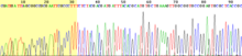
In this sequencer four different vessels are employed, each containing only of the four dideoxyribonucleotides; the incorporation of the chain terminating nucleotides by the DNA polymerase in a random position results in a series of related DNA fragments, of different sizes, that terminate with a given dideoxiribonucleotide.
The fragments are then size-separated by electrophoresis in a slab polyacrylamide gel, or more commonly now, in a narrow glass tube (capillary) filled with a viscous polymer.
This problem has been significantly reduced with the introduction of new enzymes and dyes that minimize incorporation variability.
This is changing rapidly due to the increasing cost-effectiveness of second- and third-generation systems from Illumina, 454, ABI, Helicos, and Dover.
In the array-based method (commercialized by 454 Life Sciences), single-stranded DNA is annealed to beads and amplified via EmPCR.
These DNA-bound beads are then placed into wells on a fiber-optic chip along with enzymes which produce light in the presence of ATP.
Addition of one (or more) nucleotide(s) results in a reaction that generates a light signal that is recorded by the CCD camera in the instrument.
Determining part of a protein's amino-acid sequence (often one end) by one of the above methods may be sufficient to identify a clone carrying this gene.
In many cases the assembly is not uniquely specified; depending on which enzyme acts, one of several different units may be incorporated.
Methods for the structure determination of oligosaccharides and polysaccharides include NMR spectroscopy and methylation analysis.