Siderophore
Siderophores (Greek: "iron carrier") are small, high-affinity iron-chelating compounds that are secreted by microorganisms such as bacteria and fungi.
In most aerobic environments, such as the soil or sea, iron exists in the ferric (Fe3+) state, which tends to form insoluble rust-like solids.
[7] Microbes release siderophores to scavenge iron from these mineral phases by formation of soluble Fe3+ complexes that can be taken up by active transport mechanisms.
The strict homeostasis of iron leads to a free concentration of about 10−24 mol L−1,[11] hence there are great evolutionary pressures put on pathogenic bacteria to obtain this metal.
For example, the anthrax pathogen Bacillus anthracis releases two siderophores, bacillibactin and petrobactin, to scavenge ferric ion from iron containing proteins.
[11] Because of this property, they have attracted interest from medical science in metal chelation therapy, with the siderophore desferrioxamine B gaining widespread use in treatments for iron poisoning and thalassemia.
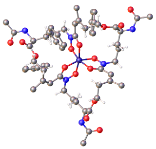
Siderophores usually form a stable, hexadentate, octahedral complex preferentially with Fe3+ compared to other naturally occurring abundant metal ions, although if there are fewer than six donor atoms water can also coordinate.
In bacteria, Fe2+-dependent repressors bind to DNA upstream to genes involved in siderophore production at high intracellular iron concentrations.
[3][8][16][24] Once in the cytoplasm of the cell, the Fe3+-siderophore complex is usually reduced to Fe2+ to release the iron, especially in the case of "weaker" siderophore ligands such as hydroxamates and carboxylates.
Under such conditions graminaceous plants (grasses, cereals and rice) secrete phytosiderophores into the soil,[25] a typical example being deoxymugineic acid.
Phytosiderophores have a different structure to those of fungal and bacterial siderophores having two α-aminocarboxylate binding centres, together with a single α-hydroxycarboxylate unit.
These mutations give an evolutionary advantage because the bacterium can benefit from siderophore production without suffering the energy cost.
This study focused on the construction, modeling, and dynamic simulation of PVD biosynthesis,[32] a virulence factor, through a systemic approach.
This approach considers that the metabolic pathway of PVD synthesis is regulated by the phenomenon of quorum-sensing (QS), a cellular communication system that allows bacteria to coordinate their behavior based on their population density.
The study showed that as bacterial growth increases, so does the extracellular concentration of QS signaling molecules, thus emulating the natural behavior of P. aeruginosa PAO1.
This group of siderophores consist of cyclic hexapeptides and consequently are highly resistant to environmental degradation associated with the wide range of hydrolytic enzymes that are present in humic soil.
Under such conditions organisms that produce hydroxamate siderophores have an advantage due to the extreme acid stability of these molecules.
[37] In contrast to most fresh-water sources, iron levels in surface sea-water are extremely low (1 nM to 1 μM in the upper 200 m) and much lower than those of V, Cr, Co, Ni, Cu and Zn.
The dilute nature of the pelagic marine environment promotes large diffusive losses and renders the efficiency of the normal siderophore-based iron uptake strategies problematic.
The presence of the fatty acyl chain renders the molecules with a high surface activity and an ability to form micelles.
Because of this continual risk of bacterial and fungal invasion, animals have developed a number of lines of defence based on immunological strategies, the complement system, the production of iron–siderophore binding proteins and the general "withdrawal" of iron.
Under normal conditions it is about 25–40% saturated, which means that any freely available iron in the serum will be immediately scavenged – thus preventing microbial growth.
In addition to these two classes of iron-binding proteins, a hormone, hepcidin, is involved in controlling the release of iron from absorptive enterocytes, iron-storing hepatocytes and macrophages.
Siderochelin is a member of the lipocalin family of proteins, which while diverse in sequence, displays a highly conserved structural fold, an 8-stranded antiparallel β-barrel that forms a binding site with several adjacent β-strands.
Siderocalin (lipocalin 2) has 3 positively charged residues also located in the hydrophobic pocket, and these create a high affinity binding site for iron(III)–enterobactin.
Poaceae (grasses) including agriculturally important species such as barley and wheat are able to efficiently sequester iron by releasing phytosiderophores via their root into the surrounding soil rhizosphere.
All of these compounds are produced by rhizospheric bacterial strains, which have simple nutritional requirements, and are found in nature in soils, foliage, fresh water, sediments, and seawater.
Examples include aluminium,[2][23][54][56] gallium,[2][23][54][56] chromium,[23][54] copper,[23][54][56] zinc,[23][56] lead,[23] manganese,[23] cadmium,[23] vanadium,[23] zirconium,[57] indium,[23][56] plutonium,[58] berkelium, californium,[59] and uranium.
[58] Alternative means of assimilating iron are surface reduction, lowering of pH, utilization of heme, or extraction of protein-complexed metal.
[2] Recent data suggest that iron-chelating molecules with similar properties to siderophores, were produced by marine bacteria under phosphate limiting growth condition.