Sphenoid wing meningioma
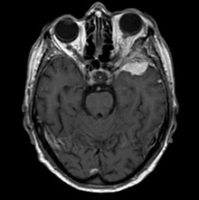
Sphenoid wing meningiomas are diagnosed by the combination of suggestive symptoms from the history and physical and neuroimaging by magnetic resonance imaging (MRI) or computer averaged tomography (CT).
Proptosis, or anterior displacement of the eye, and palpebral swelling may also occur when the tumor impinges on the cavernous sinus by blocking venous return and leading to congestion.
En plaque meningiomas characteristically lead to slowly increasing proptosis with the eye angled downward.
Angiography looking for signs like stretched arteries may be used to supplement evaluation of vascular involvement and to determine whether embolization would be helpful if surgery is being considered.
Histological factors that increase the grade include a high number of mitotic figures, necrosis and local invasion.
If surgery is done and the entire tumor cannot be removed, then external beam radiation helps reduce recurrence of the growth.
In fact, surgery followed by radiation at recurrence provided excellent tumor control in cases where gross-total resection cannot be achieved.
Untreated, one small series showed survival rates ranging from five to over twenty years, though most had unilateral blindness as well as paresis of extraocular movements.