Mitosis
This is an accepted version of this page Mitosis (/maɪˈtoʊsɪs/) is a part of the cell cycle in which replicated chromosomes are separated into two new nuclei.
[7] Other errors during mitosis can induce mitotic catastrophe, apoptosis (programmed cell death) or cause mutations.
Prokaryotes, bacteria and archaea which lack a true nucleus, divide by a different process called binary fission.
[20][21][22] The term "mitosis", coined by Walther Flemming in 1882,[23] is derived from the Greek word μίτος (mitos, "warp thread").
The genome is composed of a number of chromosomes—complexes of tightly coiled DNA that contain genetic information vital for proper cell function.
[39]: 58–67 During prophase, which occurs after G2 interphase, the cell prepares to divide by tightly condensing its chromosomes and initiating mitotic spindle formation.
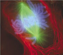
At the onset of prophase, chromatin fibers condense into discrete chromosomes that are typically visible at high magnification through a light microscope.
[43] Close to the nucleus of an animal cell are structures called centrosomes, consisting of a pair of centrioles surrounded by a loose collection of proteins.
Although centrosomes help organize microtubule assembly, they are not essential for the formation of the spindle apparatus, since they are absent from plants,[38] and are not absolutely required for animal cell mitosis.
Fungi and some protists, such as algae or trichomonads, undergo a variation called closed mitosis where the spindle forms inside the nucleus, or the microtubules penetrate the intact nuclear envelope.
[50] When a microtubule connects with the kinetochore, the motor activates, using energy from ATP to "crawl" up the tube toward the originating centrosome.
This motor activity, coupled with polymerisation and depolymerisation of microtubules, provides the pulling force necessary to later separate the chromosome's two chromatids.
[50] After the microtubules have located and attached to the kinetochores in prometaphase, the two centrosomes begin pulling the chromosomes towards opposite ends of the cell.
The resulting tension causes the chromosomes to align along the metaphase plate at the equatorial plane, an imaginary line that is centrally located between the two centrosomes (at approximately the midline of the cell).
[52] Shortening of the kinetochore microtubules pulls the newly formed daughter chromosomes to opposite ends of the cell.
The new envelope forms around each set of separated daughter chromosomes (though the membrane does not enclose the centrosomes) and the nucleolus reappears.
Even in animals, cytokinesis and mitosis may occur independently, for instance during certain stages of fruit fly embryonic development.
[9] Nuclear division takes place only in cells of organisms of the eukaryotic domain, as bacteria and archaea have no nucleus.
[70] Endomitosis is a variant of endoreduplication in which cells replicate their chromosomes during S phase and enter, but prematurely terminate, mitosis.
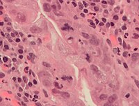
[citation needed] For example, lag-type mitosis (non-attached condensed chromatin in the area of the mitotic figure) indicates high risk human papillomavirus infection-related Cervical cancer.
[citation needed] In order to improve the reproducibility and accuracy of the mitotic count, automated image analysis using deep learning-based algorithms have been proposed.
[77][78][79] In epithelia and epidermis, an efficient rounding process is correlated with proper mitotic spindle alignment and subsequent correct positioning of daughter cells.
[78][79][80][81] Moreover, researchers have found that if rounding is heavily suppressed it may result in spindle defects, primarily pole splitting and failure to efficiently capture chromosomes.
[82] Therefore, mitotic cell rounding is thought to play a protective role in ensuring accurate mitosis.
[86][87][88] The generation of intracellular pressure is particularly critical under confinement, such as would be important in a tissue scenario, where outward forces must be produced to round up against surrounding cells and/or the extracellular matrix.
Generation of pressure is dependent on formin-mediated F-actin nucleation[88] and Rho kinase (ROCK)-mediated myosin II contraction,[84][86][88] both of which are governed upstream by signaling pathways RhoA and ECT2[84][85] through the activity of Cdk1.
[88] Due to its importance in mitosis, the molecular components and dynamics of the mitotic actomyosin cortex is an area of active research.
[89] Mutations in genes encoding enzymes employed in recombination cause cells to have increased sensitivity to being killed by a variety of DNA damaging agents.
[90][91][92] These findings suggest that mitotic recombination is an adaptation for repairing DNA damages including those that are potentially lethal.
In relation to the forms of mitosis, closed intranuclear pleuromitosis seems to be the most primitive type, as it is more similar to bacterial division.


a. non-dividing cells
b. nuclei preparing for division (spireme-stage)
c. dividing cells showing mitotic figures
e. pair of daughter-cells shortly after division