Timeline of tuberous sclerosis

TSC is a rare, multi-system genetic disease that can cause benign tumours to grow on the brain or other vital organs such as the kidneys, heart, eyes, lungs, and skin.

A combination of symptoms may include seizures, developmental delay, behavioural problems and skin abnormalities, as well as lung and kidney disease.

[1] Originally regarded as a rare pathological curiosity, it is now an important focus of research into tumour formation and suppression.

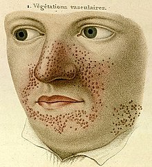
[2] In the late 19th century, notable physicians working in European teaching hospitals first described the cortical and dermatological manifestations; these early researchers have been awarded with eponyms such as "Bourneville's disease"[3] and "Pringle's adenoma sebaceum".

2012 A consensus conference was held and revised guidelines for the diagnosis and management of tuberous sclerosis were published.

Head and shoulders of a middle-aged man wearing a coat, facing his right. His receding hair is swept back and his beard is unruly, obscuring his mouth and chin.
Désiré-Magloire Bourneville first described the disease in 1880, calling it " Sclérose tubéreuse des circonvolutions cérébrales ".
A sketch consisting only of the face and shirt collar. Across the nose, the cheeks adjacent to the nose and mouth, and the chin are numerous red pimples. Above are the words "1. Végétations vasculaires."
Rayer's végétations vasculaires .
A side profile sketch of a brain, with parts indicated by a letter code. The normal convolutions of the brain are distorted in these areas, forming lumps or thickened folds.
Cerebral tuberous sclerosis showing sclerotic, hypertrophic circumvolutions.
Head and shoulders of a young lady wearing a blouse, looking slightly to her right. Her light brown hair is tied up. Her face is covered in red pimples, particularly the cheeks around her nose, the fleshy part of her nose, and her chin.
A woman with adenoma sebaceum, 1900.
The fine blood vescles seen at the back of the eye are drawn in red and dark blue on an sandy-coloured background. There are four white blobs of various sizes, one behind and one in front of the blood vescles.
Jan van der Hoeve's retinal phakoma.
Girl with TS ("epiloia"), 1926
An slice through the head, showing the cerebellum, a small portion of each temporal lobe, the ears, the orbits, and the sinuses. Bone appears white on the black background; brain, eyeballs and ears are grey.
Normal cranial CT scan.
A brightly coloured blob, roughly the shape of the head sliced horizontally, on a dark blue background. Within the head is a symmetrical pattern of blobs, having the false-colours of dark blue, cyan, green, yellow and red to indicate increasing brain activity.
Normal cranial PET scan.
A 3D skeleton of the molecular structure consisting of over around fifty small grey spheres representing carbon, linked by grey tubes. Attached to these are white spheres representing hydrogen. There are a handful of red spheres representing oxygen, and one blue sphere, which is nitrogen.
Rapamycin (sirolimus)