Coronary artery bypass surgery
Effective ways to treat chest pain (specifically, angina, a common symptom of CAD) have been sought since the beginning of the 20th century.
[1] The decision to perform surgery is informed by studies of CABG's efficacy in different patient subgroups, based on the lesions' anatomy or how well the heart is functioning.
Generally, if portions of cardiac wall are receiving less blood than normal, coronary angiography is indicated; then, lesions are identified and inform a decision to undergo PCI or CABG.
[6] CABG is generally preferred over PCI when there is a significant burden of plaque on the coronary arteries, that is extensive and complex, due to survival benefit.
Patients at risk of ongoing ischemia undergo PCI to restore blood flow and thus oxygen delivery to the struggling heart.
[7] If PCI failed to restore blood flow because of anatomical considerations or other technical problems, urgent CABG is indicated to save heart tissue.
The timing of the operation plays a role in survival: It is preferable to delay the surgery if possible (three days if the infarction affecting the total thickness of the cardiac muscle, and six hours if it does not).
[8] There are no absolute contraindications of CABG, but severe disease of other organs such as the liver or brain, limited life expectancy, and patient fragility are considered.
[8] CABG is also performed when a patient is to undergo another cardiac surgical procedure, most commonly for valve disease, and angiography reveals a significant lesion of the coronary arteries.
The choice of method is still a matter of debate, but it is clear that in the presence of complex lesions, significant left main disease, or diabetes, CABG yields better long-term survival and outcomes.
Studies published in 2023 show that CABG in patients with left main disease is associated with lower mortality and fewer adverse events compared to PCI.
The examination typically includes a chest X-ray to check the lungs, a complete blood count, and kidney and liver function tests.
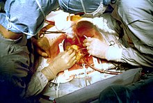
[21] An incision in the sternum is made while vessels are being harvested, either from the arms or chest or from the leg, usually from the internal mammary artery or the saphenous vein.
Deoxygenated blood arriving to the heart from veins is forwarded to the CPB machine to get oxygenated, then delivered to the aorta to keep the rest of the body saturated.
[21][23] Off-pump coronary artery bypass (OPCAB) surgery avoids using the CPB machine by stabilizing small segments of the heart at a time.
The surgical team and anesthesiologists must coordinate and take great care to not manipulate the heart too much, lest they compromise the stability of blood flow.
[30] Angiotensin-converting enzyme (ACE) inhibitors and angiotensin receptor blockers (ARBs) are used to control blood pressure, especially in patients with low cardiac function (<40%).
[35] However, the use of bilateral mammary artery in patients of younger age and those without specific comorbidities (diabetes, obesity, steroid use) can provide excellent long-term survival and quality of life.
The ITAs are advantageous because of their endothelial cells, which produce endothelium-derived relaxing factor and prostacyclin, protecting the artery from atherosclerosis and thus stenosis or occlusion.
[10] The most common complications of CABG are postoperative bleeding, heart failure, atrial fibrillation (a form of arrhythmia), stroke, kidney dysfunction, and infection of the wound near the sternum.
According to its severity, LCOS is treated with inotropes, an intra-aortic balloon pump (IABP), optimization of pre-load and afterload, or correction of blood gauzes and electrolytes.
[47] Arrhythmias can also occur, most-commonly atrial fibrillation (incidence of 20–40%) that is treated with correcting electrolyte balance, and rate and rhythm control.
[39] In the early 20th century, surgical interventions aiming to relieve angina and prevent death were either sympathectomy — a cut on the sympathetic chain that supplies the heart—or pericardial abrasion, with the hope adhesions would create significant collateral circulation.
[50] French surgeon Alexis Carrel was the first to anastomose a vessel—a branch of the carotid artery—to a native artery in the heart of a dog, but the experiment could not be reproduced.
[50] The development of coronary angiography in 1962 by Mason Sones helped medical doctors to identify patients in need of operation, and which native heart vessels should be bypassed.
[52] In 1964, Soviet cardiac surgeon Vasilii Kolesov performed the first successful internal thoracic artery–coronary artery anastomosis, followed by Michael DeBakey in the United States.
[53] The modern era of the CABG began in 1964 when Soviet cardiac surgeon Vasilii Kolesov performed the first successful internal thoracic artery–coronary artery anastomosis.
[54] In the late 1960s, after the work of René Favaloro, the operation was performed in only a few centers, but was anticipated to more broadly change the outcome of coronary artery disease.
The introduction of percutaneous coronary intervention (PCI) did not obsolesce CABG; rates of both procedures continued to increase, but PCIs grew more rapidly.
In 1971, Carpentier introduced the use of the radial artery, which was initially prone to failure, but the development of harvesting techniques in the following 20 years significantly improved patency.