Vascular anomaly
A vascular anomaly is any of a range of lesions from a simple birthmark to a large tumor that may be disfiguring.
Some vascular anomalies are congenital, others appear within weeks to years after birth, and others are acquired by trauma or during pregnancy.
[2] Vascular anomalies can occur throughout the whole body, but in 60% of patients they are localized in the head and neck region.
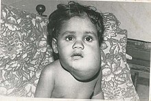
[3] Vascular anomalies can present in various ways: when situated deep below the skin, they appear blue, and are often called cavernous.
Historically, vascular anomalies have been labeled with descriptive terms, according to the food they resembled (port wine, strawberry, cherry, salmon patch).
[4] However, in 1982, Mulliken introduced a classification that replaced these descriptive terms and gave direction to the management of various vascular anomalies.
[3] Vascular tumors are characterized by an overgrowth of normal vessels, which show increased endothelial proliferation.
Since 90% of IH is small, localized and asymptomatic, treatment mainly consists of observation and awaiting until involution is complete.
IH can be treated with corticosteroids, which accelerate involution: in 95% of patients, growth is stabilized and 75% of tumors decrease in size.
Systemic corticosteroids may cause a number of side-effects and are only used in problematic IH, which is too large to treat with intralesional injections.
However, patients may require intervention during childhood, because 50% of IH leave residual fibrofatty tissue, redundant skin, or damaged structures after involution.
Waiting until involution is completed, ensures that the least amount of fibro fatty residuum and excess skin is resected, giving the smallest possible scar.
Unlike IH, CH is more common in the extremities, has an equal sex distribution, and is solitary, with an average diameter of 5 cm.
The rapidly involuting congenital hemangioma, RICH, presents at birth as a solitary raised tumor with a central depression, scar, or ulceration surrounded by a rim of pallor.
[6] The non-involuting congenital hemangioma, NICH, presents as a solitary, well-circumscribed reddish-pink to purple plaque with central telangiectasia and hypopigmented rim.
It persists into late childhood and can even mimic a vascular malformation by growing commensurately with the child.
Resection may be indicated to improve the appearance of the affected area, as long as the surgical scar is less noticeable than the lesion.
The use of corticosteroids leads to accelerated regression in 30%, stabilization of growth in 40%, lightening of color and softening of the tumor.
Other therapeutic options are embolization and pulsed-dye laser, which improves residual telangiectasias in RICH and in NICH.
[citation needed] Kaposiform hemangioendothelioma (KHE) is a rare vascular neoplasm that is locally aggressive but without metastatic potential.
Although lesions occur solitary, they often involve large areas of the body, such as the head/neck region (40%), trunk (30%), or extremity (30%).
Although complete surgical removal with a large margin has the best reported outcome, it is usually not done because of the risk of bleeding, extensiveness, and the anatomic site of the lesion.
[citation needed] A pyogenic granuloma, is a small benign vascular tumor that primarily involves the skin (88.2%) and mucous membranes.
[6] Pyogenic granuloma appears as a red macule that grows rapidly, turns into a papule and eventually becomes pedunculated, being attached to a narrow stalk.
Microscopically, pyogenic granulomas are characterized by vascular proliferation amidst granulation tissue and chronic inflammatory infiltrate.
[6] This vascular tumor is twice as common in males as in females and 25% of lesions seem to be associated with trauma, an underlying cutaneous condition, pregnancy, hormonal alterations and medications.
Lesions involving the reticular dermis, may be out of the reach of pulsed-dye laser, cautery or shave excision and therefore have a recurrence rate of 43.5%.