MRI artifact
Phase-encoded sampling takes several seconds, or even minutes, owing to the collection of all the k-space lines to enable Fourier analysis.
Major physiological movements are of millisecond to seconds duration and thus too slow to affect frequency-encoded sampling, but they have a pronounced effect in the phase-encoding direction.
High velocity flow causes the protons entering the image to be removed from it by the time the 180-degree pulse is administered.
[1] Determination of the artifact can be made by swapping the phase- and frequency-encoding gradients and examining the resultant shift of fat tissue.
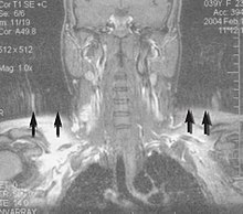
It appears as multiple, regularly spaced parallel bands of alternating bright and dark signal that slowly fade with distance (Fig.
[1] Methods employed to correct Gibbs artifact include filtering the k-space data prior to Fourier transform, increasing the matrix size for a given field of view, the Gegenbauer reconstruction and Bayesian approach.
Similarly, for inversion recovery pulses, and other T1-dependent methods, will suffer from signal intensity errors and generally lower T1 weighting.
[9] But as the main magnetic field is increased, these wavelengths become the same or smaller than the regions of the body being imaged, resulting in flip angle inhomogeneity.
[1] Zero line and star artifacts are due to system noise or any cause of RF pollution within the room (Faraday cage).
[1] Further from the coil the signal strength drops rapidly due to the attenuation with a loss of image brightness and significant shading to the uniformity.
Retrospective techniques using the pilot tone are able to increase the level of detail and reduce blurring in free-breathing radial images.
[16] The TAMER Method utilizes the SENSE forward model (described below) that has been modified to include the effects of motion in a 2D multi-shot imaging sequence.
Note: the following modified SENSE model is described in detail in Melissa Haskell's doctoral dissertation, Retrospective Motion Correction for Magnetic Resonance Imaging.
In the effort to speed up and simplify computations, the TAMER method separates the vector of image voxel values,
As described in the paper Targeted Motion Estimation and Reduction (TAMER): Consistency Based Motion Mitigation for MRI using a Reduced Model Joint Optimization, as part of the IEEE Transactions on Medical Imaging Journal, the TAMER algorithm converges fastest when choosing target voxels that are highly coupled.
[17] Joint Optimization Reduced Model Search: We now have the initial target voxels, motion estimate, and coil groupings.
repeat the following: Advantages: Disadvantages: In recent years, neural networks have generated a great deal of interest by outperforming traditional methods[18] on longstanding problems across many fields.
This serves two purposes: First, it allows the CNN to perform backpropagation and update its model weights by using a mean square error loss function comparing the difference between
Using a CNN effectively allows us to bypass the second stage of TAMER by skipping the joint parameter search.
The NAMER technique has shown itself to be very effective in correcting for rigid motion artifacts, and converges much faster than other methods including TAMER.
B1 inhomogeneity due to constructive or destructive interference from the permittivity of body tissue can be mitigated using external objects with high dielectric constants and low conductivity.
The combination of high dielectric constant and having low conductivity allows the cushion to alter the phase of the RF standing waves and has been shown to reduce signal loss due to B1 inhomogeneity.
For anatomical studies using the FLASH sequence that can be performed with one transmit and receive coil, this method can be used to reduce B1 inhomogeneity artifacts.
However, the method would not be suitable for exams under strict time constraints, since the user first needs to perform flip angle optimization.
Overall, when this method is used with the correct selection of RF pulses and optimized for a low power deposition, the artifacts from B1 inhomogeneity can be greatly reduced.
[32] This method focuses on inhomogeneity along the axial, or z axis, direction since it is the most dominant in terms of poor homogeneity and least sample dependent.
[32] This method is advantageous for reducing artifacts at the source, particularly when accurate flip angle is critical and for increasing signal to noise ratio.
This correction method works by removing the source of the problem and applying pulses that will not generate flip angle errors.
The subsequent image enhancement can be processed with shorter scan times for higher throughput and plausible earlier detection.
DREAM can be used to acquire a 2D B1 map in 130 ms, making it insensitive to motion and feasible for scans that require breath holds, such as cardiac imaging.